. 2007 Jun 20;26(12):2843-55.
doi: 10.1038/sj.emboj.7601724. Epub 2007 May 24.

p140Cap protein suppresses tumour cell properties, regulating Csk and Src kinase activity

Affiliations

p140Cap protein suppresses tumour cell properties, regulating Csk and Src kinase activity

Paola Di Stefano et al. EMBO J. .

Abstract

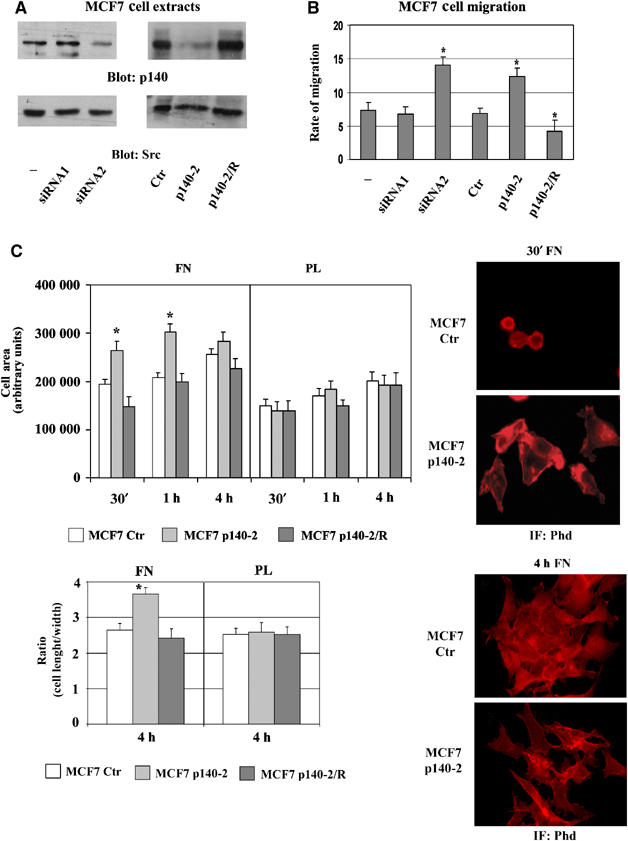
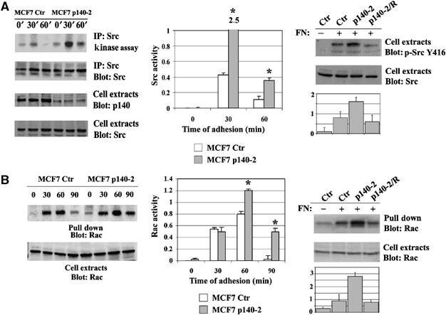
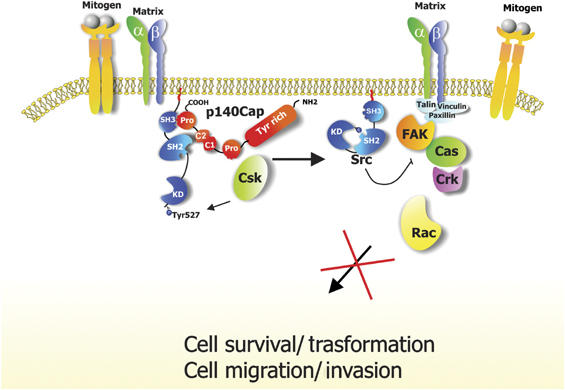
We recently identified p140Cap as a novel adaptor protein, expressed in epithelial-rich tissues and phosphorylated upon cell matrix adhesion and growth factor treatment. Here, we characterise p140Cap as a novel Src-binding protein, which regulates Src activation via C-terminal Src kinase (Csk). p140Cap silencing increases cell spreading, migration rate and Src kinase activity. Accordingly, increased expression of p140Cap activates Csk, leading to inhibition of Src and downstream signalling as well as of cell motility and invasion. Moreover, cell proliferation and "in vivo" breast cancer cell growth are strongly impaired by high levels of p140Cap, providing the first evidence that p140Cap is a novel negative regulator of tumour growth.


Figures

Figure 1
Figure 1
p140Cap silencing enhances cells spreading and migration. (A) p140Cap expression was evaluated by Western blotting on SDS–PAGE in extracts of MCF7 p140-2, MCF7 Ctr, MCF7 cells transiently transfected with control (−), scrambled (siRNA1) or human p140Cap (siRNA2) siRNAs and in MCF7 p140-2/R, transiently transfected with murine pcDNA3.1 Myc-p140Cap to reconstitute p140Cap expression. The same blot was re-probed with antibodies to Src kinase. (B) Cells as in (A) were tested for migration in Transwell assays. Cells were seeded on the upper surface, left to migrate for 9 h in the presence or the absence of HGF (25 U/ml), then fixed, stained and counted. Numbers on the y-axes represent the rate of migration as the ratio between the number of cells migrated in response to the presence and the absence of HGF. (C) MCF7 Ctr, p140-2 and p140-2/R cells were plated on FN and on PL for the indicated times, fixed and stained with phalloidin (Phd). Left upper panel, the histogram represents the mean cell area for each time point in arbitrary units. The area of attached cells was calculated by Metamorph Software in 20 random fields (200 cells) at a magnification of × 60. Left lower panel, the histogram represents the mean ratio between cell length and width as a measure of cell shape at 4 h of adhesion on at least 200 cells. Right panels, representative fields at 30 min (upper) and 4 h (lower) of adhesion are shown at × 60 magnification. The results are representative of six independent experiments (*P<0.05).
Figure 2
Figure 2
p140Cap silencing enhances Src and Rac activities. (A) Left panel, MCF7 Ctr or p140-2 cells were plated on FN for 30 and 60 min or kept in suspension (0). Right panel, MCF7 Ctr, p140-2 and p140-2/R were plated on FN for 60 min. Src kinase assay was performed as described in Material and Methods using Enolase as Src substrate. The amount of p140Cap and Src in cell extracts was evaluated by Western blot with specific antibodies. (B) Left panel, MCF7 Ctr or p140-2 cells were plated on FN for 30, 60 and 90 min or kept in suspension (0). Right panel, MCF7 Ctr, p140-2 and p140-2/R were plated on FN for 60 min. Activated Rac was pulled down from 1.5 mg of protein extract with the CRIB domain of PAK and detected by Western blot with anti-Rac mAb (upper panel). Total amount of Rac protein in cell extracts is shown in the lower panel. The histograms show the ratio between active and total protein levels in arbitrary units (*P<0.05).
Figure 3
Figure 3
p140Cap overexpression inhibits cells spreading, migration and invasion. (A) MCF7-Mock, p140/P9 and p140/P23 cells were plated on FN for the indicated times in absence (left panel) or in presence of serum (right panel), fixed and stained with Phd. The histogram represents the mean cell area for each time point in arbitrary units. The area of attached cells was calculated by Metamorph Software in 20 random fields (200 cells) at a magnification of × 60. (B) MCF7-Mock (a–c) and p140/P9 (d–f) cells as in (A) (Right panel) were fixed and stained with Diff Quick kit and photographed at 20X magnification. (C) MCF7-Mock, p140/P9 and p140/P23 cells were tested for migration and invasion in Transwell assays. Cells were left to migrate for 9 h in the presence or the absence of HGF (25 U/ml), then fixed, stained and counted. For invasion, transwells were coated with Matrigel and cells left to invade for 48 h. Numbers on the y-axes represent the fold increase in migration in response to HGF compared to nonstimulated cells. The mean values were calculated on six independent experiments (*P<0.05).
Figure 4
Figure 4
p140Cap overexpression compromises integrin-dependent Src activation and downstream signalling. MCF7-Mock and p140/P9 were plated on FN or kept in suspension (S) for 30 min and extracted. (A) Left panel, Src kinase assay on Enolase was performed as described in Material and Methods. Right panel, in the same extracts, Src phosphorylation on tyrosine 416 was evaluated using phosphospecific antibodies (Y416). The total amount of Src was evaluated by Western blot. (B) FAK phosphorylation was evaluated in cell extracts using phosphospecific antibodies for the FAK autocatalytic tyrosine 397 (Y397) (Left panel) and FAK tyrosine 925 (Y925) (Right panel). The total amount of FAK was quantified using FAK mAbs. (C) Cell extracts from MCF7-Mock and p140/P9 plated on FN for 30 min were immunoprecipitated with Src antibodies and pre-immune serum as negative control. The amount of FAK co-immunoprecipitated with Src was detected with FAK antibody. The level of immunoprecipitated Src was quantified using Src polyclonal antibodies. (D) Cell extracts as in (A) were immunoprecipitated with anti phosphotyrosine antibodies (P-Tyr) followed by Western blot analysis with p130Cas mAbs. p130Cas was evaluated in cell extracts. (E) Cell extracts as in (A) were analysed for activated Rac as described in Figure 2B. (F) Cell extracts as in (A) were analysed for Akt phosphorylation using phosphospecific antibodies for the Akt Serine 473 Ser(473). The total amount of Akt was quantified using antibodies. The results are representative of three independent experiments. The histograms show the ratio between active or phosphorylated and total protein levels in arbitrary units (*P<0.05).
Figure 5
Figure 5
p140Cap binds directly to Src SH3 domain. (A) MCF7 cell extracts were immunoprecipitated with mAbs to p140Cap or Src. The immunoprecipitates were blotted with Src antibodies. Molecular weights are indicated on the left. (B) MCF7 cell extracts were immunoprecipitated with mAbs to p140Cap or pre-immune serum. The immunoprecipitate were blotted with anti-p140Cap polyclonal antibodies and re-probed with anti-pTyr antibodies. The results are representative of six independent experiments. (C) Upper panel, a schematic representation of full-length p140Cap domains. Lower panel, GST, GST-Src/SH2 and GST-Src/SH3 fusion proteins were used to pull down p140Cap from MCF7 cell extracts. Bound proteins were immunoblotted using p140Cap polyclonal antibodies (left) or stained with Coomassie blue (right) to quantify the loading. The results are representative of two independent experiments. (D) A total of 2 μg of purified p140Cap GST-Cap3, -4 and -5 fusion proteins (left panel) were incubated in ‘in vitro' binding assays with 2 μg of MBP-Src/SH2 or MBP-Src/SH3 proteins. Associated proteins were pulled down with Glutathione–Sepharose, eluted and immunoblotted with anti MBP antibodies. The results are representative of two independent experiments.
Figure 6
Figure 6
p140Cap inhibition of Src kinase activity is dependent on Csk activity. (A) MCF7-Mock and p140/P9 cells were plated on FN for different times or kept in suspension (S). Phosphorylation of Src Tyr-527 was detected by Western blot with specific antibodies. The same blots were re-probed with antibodies to Src. The histogram shows the ratio between Tyr-527 phosphorylation and Src protein levels in arbitrary units. The results are representative of three independent experiments. (B) Left panels, cell extracts of MCF7-Mock and p140/P9 plated on FN for 30 min, were immunoprecipitated with antibodies to Csk or pre-immune rabbit immunoglobulins (PI) and subjected to kinase assay. The amount of immunoprecipitated Csk was evaluated by Western blot with specific antibodies. Right panel, densitometric analysis of Poly-Glu-Tyr signal is reported in arbitrary units. The results are representative of two independent experiments. (C) Mock-MCF7 and p140/P9 cells were infected with GFP or kinase-negative mutant of Csk (Csk-KD) recombinant adenoviruses. Infected cells were plated on FN for 30 min. Src activity was analysed by in vitro kinase assay as shown in Figure 2A. The level of Csk was detected by Western blot. Arrow indicates immunoglobulin. The results are representative of three independent experiments. (D) MCF7-Mock cell extracts were immunoprecipitated with mAbs to p140Cap, Csk or pre-immune serum (PI) as negative control. Immunocomplexes were analysed by Western blotting using antibodies to Src, Csk and p140Cap, respectively. The results are representative of four independent experiments. (E) Cell extracts from HEK293 transfected with p140Cap were immunoprecipitated with mAbs to p140Cap and GAPDH. Upper panel, Western blot was probed with recombinant Csk produced by in vitro transcription/translation followed by anti-Csk antibodies. Middle and lower panels, the blots were re-probed with anti-p140Cap and GAPDH antibodies. L, lysates; MW, molecular weight.
Figure 7
Figure 7
Src-binding domain is essential for p140Cap role in biological processes. (A) Left panel, a schematic representation of full-length p140Cap protein, p140Pro and p140Delta mutants. Right panel, cell extracts of MCF7p140/P9, p140Pro and p140Delta were analysed by Western blot using Myc antibodies. The same filter was re-probed with Src-specific antibodies. (B) Extracts of HEK293 cells transiently transfected with p140FL, p140Pro and p140Delta were immunoprecipitated with Src antibodies. The immunoprecipitate were analysed by Western blot with Myc and Src antibodies. (C) The histogram represents the mean cell area for MCF7-Mock, p140/P9, p140Pro and p140Delta cells plated on FN for the indicated times, calculated as described in Figure 1C. (D) Left panel, the same cells as in (C) were induced to migrate and to invade as described in Figure 3C. (E) Extracts of MCF7-Mock and p140Delta cells plated on FN for 30 min or kept in suspension (S) were tested for in vitro Src kinase assay as shown in Figure 2A (left panel) or for Rac activation as shown in Figure 2B. The results are representative of three independent experiments (*P<0.05).
Figure 8
Figure 8
p140Cap regulates anchorage-independent and in vivo tumour growth. (A) Upper panel, expression of p140Cap was evaluated in extracts of MCF7, T47D, and MDA-MB-231 and MDA-MB-435 breast cancer cells by Western blot with p140Cap antibodies. The blot was re-probed with Src antibodies. Lower panel, MDA-MB-231 cells stably transfected with p140Cap-Myc were analysed by Western blot with anti-Myc tag antibodies. Cell population P12 and P16 were selected for further experiments. (B) MDA-MB-231 Mock, p140/P12 and p140/P16 cells were tested for their ability to migrate for 2 h (upper panel) or to invade Matrigel-coated Transwells for 12 h (lower panel) as described in Figure 3C. The mean values were calculated on five independent experiments (*P<0.05). (C) 107 MDA-MB-231 Mock, p140/P16 and p140Delta cells, transfected with the deleted mutant p140Delta, were injected subcutaneously in SCID mice. Progressively growing neoplastic masses were measured with calipers and the average value recorded as mean tumour diameter. Differences in tumour volume were evaluated with Student's t-test. (D) 105 MDA-MB-231 Mock, p140/P16 or p140Delta cells were allowed to grow in the presence of 10% FCS for 12 days. Every two days, cells were detached and counted. The mean number of cells from three separate experiments is reported on the y-axis. (E) Left panel, 105 MCF7-Mock, p140/P9 and p140Delta cells were allowed to grow for 12 days and counted as in (D) (right upper panel). Extracts of MCF7-Mock and p140/P9 cells grown for 2, 4 and 6 days were analysed by Western blot with cyclin D1 and actin antibodies. Right lower panel, extracts of MCF7-Mock and p140/P9 cells starved for 24 h and treated for 30 min with 20% FCS or left untreated, were analysed for Src kinase assay as described in Figure 2A. The amount of Src in immunoprecipitates was evaluated by Western blot. The results are representative of three independent experiments.
Figure 9
Figure 9
Schematic summary of the signalling pathways affected by p140Cap. Upon cell matrix adhesion or mitogen stimulus, Src is activated and recruits additional transducing molecules such as FAK and p130Cas, leading to cell migration and invasion and regulation of cell growth and transformation. p140Cap participates to these processes by activating Csk, inhibiting Src activity and downstream signalling, thus leading to impaired cell spreading, motility, invasion and growth.

References

    1. Avizienyte E, Wyke AW, Jones RJ, McLean GW, Westhoff MA, Brunton VG, Frame MC (2002) Src-induced de-regulation of E-cadherin in colon cancer cells requires integrin signalling. Nat Cell Biol 4: 632–638 - PubMed
    1. Bon G, Folgiero V, Bossi G, Felicioni L, Marchetti A, Sacchi A, Falcioni R (2006) Loss of beta4 integrin subunit reduces the tumorigenicity of MCF7 mammary cells and causes apoptosis upon hormone deprivation. Clin Cancer Res 12: 3280–3287 - PubMed
    1. Bouton AH, Riggins RB, Bruce-Staskal PJ (2001) Functions of the adapter protein Cas: signal convergence and the determination of cellular responses. Oncogene 20: 6448–6458 - PubMed
    1. Boyer B, Roche S, Denoyelle M, Thiery JP (1997) Src and Ras are involved in separate pathways in epithelial cell scattering. EMBO J 16: 5904–5913 - PMC - PubMed
    1. Brummelkamp TR, Bernards R, Agami R (2002) A system for stable expression of short interfering RNAs in mammalian cells. Science 296: 550–553 - PubMed

Publication types

MeSH terms