. 2007 Jun;117(6):1490-501.
doi: 10.1172/JCI29882. Epub 2007 May 24.

Pancreas-specific RelA/p65 truncation increases susceptibility of acini to inflammation-associated cell death following cerulein pancreatitis

Affiliations

Pancreas-specific RelA/p65 truncation increases susceptibility of acini to inflammation-associated cell death following cerulein pancreatitis

Hana Algül et al. J Clin Invest. 2007 Jun.

Abstract

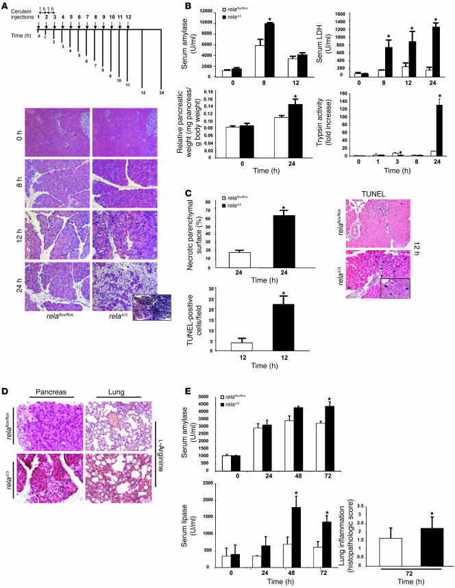
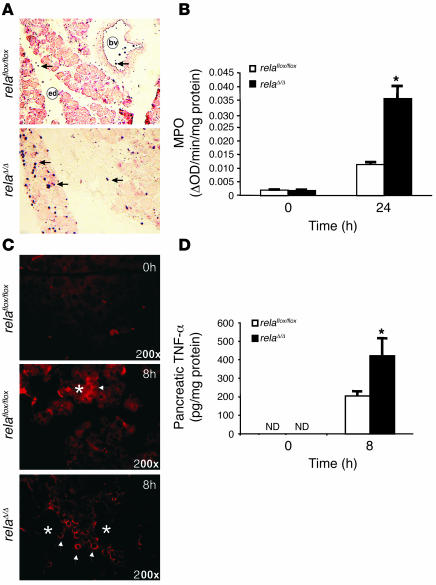
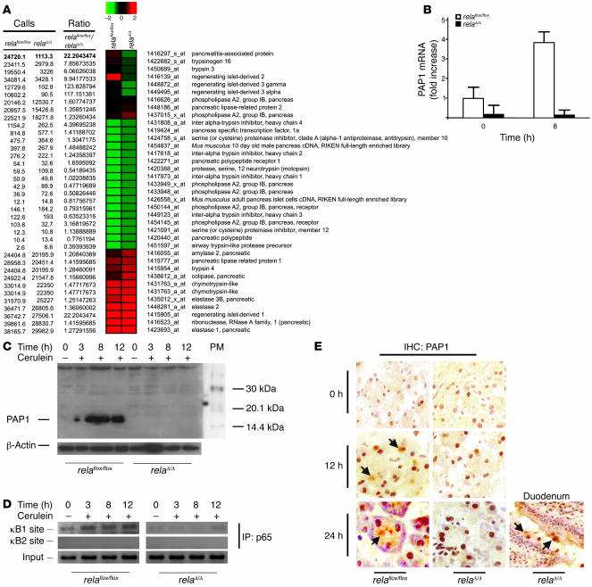
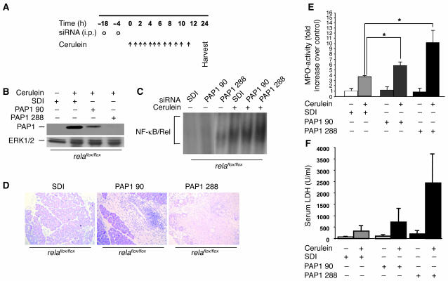
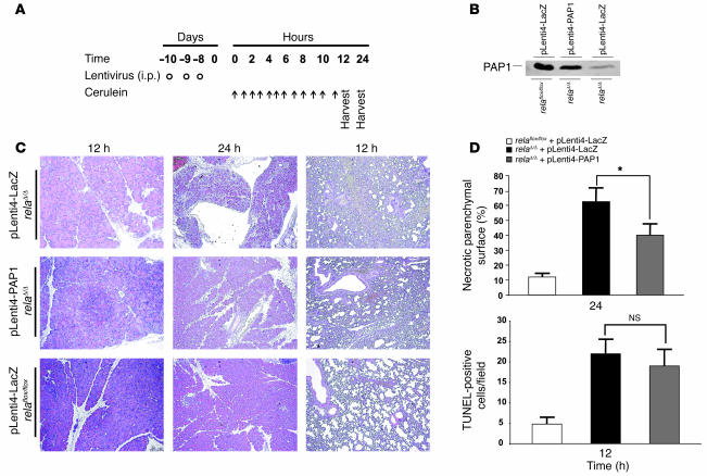
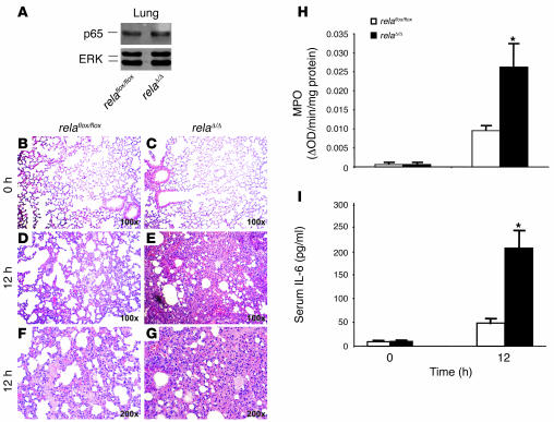
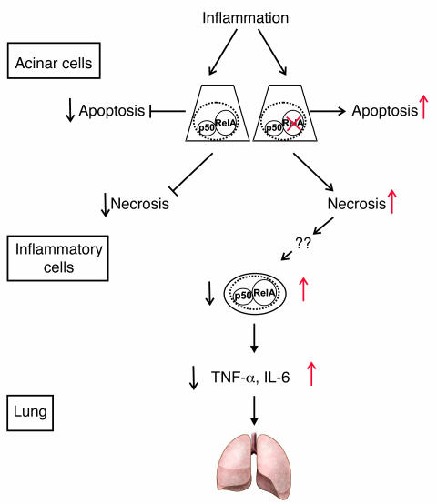
Activation of the transcription factor NF-kappaB/Rel has been shown to be involved in inflammatory disease. Here we studied the role of RelA/p65, the main transactivating subunit, during acute pancreatitis using a Cre-loxP strategy. Selective truncation of the rela gene in pancreatic exocrine cells led to both severe injury of the acinar cells and systemic complications including lung and liver damage. Our data demonstrated that expression and induction of the protective pancreas-specific acute phase protein pancreatitis-associated protein 1 (PAP1) depended on RelA/p65. Lentiviral gene transfer of PAP1 cDNA reduced the extent of necrosis and infiltration in the pancreata of mice with selective truncation of RelA/p65. These results provide in vivo evidence for RelA/p65 protection of acinar cell death via upregulation of PAP1. Moreover, our data underscore the pancreas-specific role of NF-kappaB/Rel and suggest multidimensional roles of NF-kappaB/Rel in different cells and contexts during inflammation.


Figures

Figure 1
Figure 1. Pancreas-specific truncation of RelA/p65 using a Cre-loxP system.
(A) Two identically oriented loxP sites (triangles) flank exons 7 and 10 of the rela gene. loxPmod, modified loxP site. (B) Recombination of genomic DNA from the pancreata of relaΔ/Δ mice was detected by Southern blot analysis using a probe external to the 5′ end of the targeting construct. No recombination was detected in the other organs. (C) Deletion of rela (exons 7–10) in the mouse pancreas was demonstrated at the protein level by Western blot of isolated acini from mice with the floxed allele in the presence or absence of Cre as indicated. In contrast to relaflox/flox mice, relaΔ/Δ mice display a truncated form of RelA/p65 (Δp65). Pancreas protein extracts (40 μg) were analyzed using antibodies against p50, IκBα, and β-actin (as a loading control). The protein marker (PM) indicates the size of detected protein bands.
Figure 2
Figure 2. Inhibition of nuclear translocation of RelA/p65 in the pancreas following cerulein stimulation.
(A) relaflox/flox and relaΔ/Δ mice were injected i.p. with 50 μg/kg cerulein at hourly intervals. Pancreatic nuclear protein extracts (10 μg) at the indicated time points were subjected to gel retardation assays with an NF-κB consensus probe. (B) Kinetics of IκBα degradation in relaflox/flox and relaΔ/Δ mice were assessed by Western blot analyzing the protein content of 40 μg of pancreatic whole-cell protein lysates using an antibody against IκBα. Phosphorylated IκBα (p-IκBα; Ser32) was monitored by Western blotting using a phosphospecific antibody.
Figure 3
Figure 3. Pancreas-specific truncation of RelA/p65 exacerbates AP.
(A) relaflox/flox and relaΔ/Δ mice were given 8 hourly i.p. injections of cerulein (50 μg/kg) and sacrificed 8, 12, or 24 hours after the first injection. Histological sections of relaflox/flox and relaΔ/Δ mice were analyzed at the indicated time points. Note the increased vacuolization, morphologically apoptotic cells, ghost cells, edema, infiltration, and massive necrosis in the relaΔ/Δ pancreata. (B) Pancreatic injury was determined by measuring amylase and LDH enzyme activity in serum. Total tissue homogenates were obtained from pancreata of cerulein-injected mice at the indicated time points and subjected to trypsin activity analysis. Pancreatic edema was determined indirectly by increase in pancreatic weight. (C) H&E-stained pancreas sections from relaflox/flox and relaΔ/Δ mice 24 hours after cerulein-induced inflammation were used to measure and quantify necrotic parenchymal surface area. TUNEL assay results are expressed as the apoptotic index of pancreata from mice with AP. Apoptotic cells exhibited black nuclei. (D) l-Arginine–induced pancreatitis was evaluated 72 hours after induction. Pancreata and lungs were removed for morphological analysis by H&E. Note the appearance of focal necrosis in the pancreata of both groups. (E) Serum was removed for amylase and lipase evaluation at the indicated time points. Note the significant release of amylase and lipase into the serum in relaΔ/Δ mice. Lung inflammation was evaluated as described in Methods. Values are mean ± SD for independent animals (n = 5). *P < 0.05 versus relaflox/flox. Original magnification, ×50 (A, inset); ×200 (A and D); ×100 (C, inset); ×100 (C).
Figure 4
Figure 4. Analysis of MAPK modules during cerulein-induced pancreatitis.
Pancreata were removed at the indicated time points. Whole-cell lysates (30 μg) were blotted against phosphorylated p42, p44, p38, p46, and p54 and their respective unphosphorylated proteins.
Figure 5
Figure 5. Increased infiltration and TNF-α production in the pancreata of relaΔ/Δ mice.
(A) Mononuclear infiltration into the pancreas was visualized by immunohistochemical detection of granulocytes using a Gr-1 antibody. bv, blood vessel; ed, edema. Original magnification, ×100. (B) Pancreatic MPO activity was measured in the pancreata of relaflox/flox and relaΔ/Δ mice. (C) Infiltrating cells produced and released TNF-α. Pancreata from cerulein-treated relaflox/flox and relaΔ/Δ mice were stained using antibody to TNF-α. Arrowheads indicate TNF-α accumulation; asterisks indicate acinar cells. Original magnification, ×200. (D) TNF-α levels were measured by ELISA in pancreatic cell lysates from relaflox/flox and relaΔ/Δ mice following 8 hourly injections of 50 μg/kg cerulein. ND, not detected. Values are mean ± SD for independent animals (n = 4). *P < 0.05 versus relaflox/flox.
Figure 6
Figure 6. Impaired upregulation of murine PAP1 during AP in relaΔ/Δ mice.
(A and B) Pancreata from relaflox/flox and relaΔ/Δ mice were removed at the indicated times. (A) Total pancreatic RNA (8 μg; n = 2) was labeled and hybridized to Affymetrix MOE430A GeneChips, and pancreas-specific genes were clustered hierarchically. (B) Relative levels of PAP1 mRNA were determined by real-time PCR and expressed as mean ± SD (n = 5). (C) Pancreatic tissues were removed at the indicated times. Whole-tissue extracts were prepared and subjected to Western blot analysis using a newly generated antibody to murine PAP1. (D) Chromatin immunoprecipitation experiments were performed with relaΔ/Δ and relaflox/flox pancreatic tissue at the indicated times after stimulation with cerulein using an antibody to p65. Precipitated DNA was analyzed by PCR using primers surrounding the positions of both κB sites in the respective promoters. PCR was also performed with 2.5% of input chromatin to ensure equal loading. (E) Sections of snap-frozen pancreata and duodenum were prepared and analyzed for PAP1 expression in relaflox/flox and relaΔ/Δ mice in unstimulated pancreas and 12 and 24 hours after the first cerulein injection, respectively. Snap-frozen duodenum served as a positive control, because Paneth cells are known to express PAP1. Signals in the pancreas were localized to the apical regions of acini (arrows), typical for secretory proteins like PAP1.
Figure 7
Figure 7. Inhibition of the protective effect of PAP1 using PAP1 knockdown in vivo.
(A) Three age- and sex-matched relaflox/flox mice were injected with SDI or PAP1 siRNA (PAP1 90 and PAP1 288) twice in an 18-hour interval (circles) and then were subjected to cerulein-induced pancreatitis (arrows). (B) Pancreatic homogenates from mice as in A were obtained and immunoblotted for PAP1. Blotting for ERK1/2 protein was used as a loading control. (C) relaflox/flox mice were pretreated with specific and nonspecific siRNA as in A and subsequently injected with 1 i.p. dose of 50 μg/kg cerulein. One hour after injection, pancreatic nuclear protein extracts (10 μg) were subjected to gel retardation assays with an NF-κB consensus probe. (D) Representative H&E staining of pancreata from mice treated as in A. Note the increase in infiltration and massive necrosis in mice with PAP1 knockdown. (E and F) Pancreatic injury was measured by determining the enzyme activity of MPO in the pancreas (E) and LDH in the serum (F). Values are mean ± SD for independent animals (n = 3). *P < 0.05.
Figure 8
Figure 8. Protective effect of PAP1 on cerulein-induced pancreatitis in relaΔ/Δ mice.
(A) Lentivirus harboring the full-length cDNA of murine PAP1 was generated in HEK 293T cells and injected i.p. into relaΔ/Δ mice to express PAP1 (pLenti4-PAP1) in the pancreas (circles). Four age- and sex-matched mice were used. Littermate control mice were infected with lentivirus (pLenti4-LacZ) containing the lacZ gene. Mice were subjected to cerulein-induced pancreatitis (arrows) 7 days after infection and then were sacrificed 12 and 24 hours after the first cerulein injection. (B) Pancreatic homogenates were obtained 12 hours after the first cerulein injection and analyzed for PAP1 expression. (C) Representative H&E-stained sections of pancreata and lungs from mice treated as in A. Original magnification, ×100. (D) To assess the extent of tissue injury, necrosis and apoptosis were evaluated. Areas of necrotic parenchymal surface were measured and quantified. TUNEL assay was used to determine the apoptotic index of the pancreas. Values are mean ± SD for independent animals (n = 4). *P < 0.05.
Figure 9
Figure 9. Pancreas-specific ablation of RelA/p65 promotes systemic complications and multiorgan dysfunction syndrome.
(A) Expression of RelA/p65 in lung tissue of relaflox/floxand relaΔ/Δ mice was assessed by subjecting 20 μg of lung whole cell lysates to Western blot analysis. (BG) At the indicated times after cerulein injection, lung tissue was removed, embedded in paraffin, and stained with H&E. Higher magnification of representative H&E stains reveal marked hemorrhage and alveolar collapse in relaΔ/Δ mice compared with relaflox/flox mice. Original magnification, ×100 (BE); ×200 (F and G). (H) Lung tissue of relaflox/floxand relaΔ/Δ mice was removed at the indicated time points and used to determine MPO enzyme activity. (I) Serum concentrations of IL-6 were determined at the time points indicated. Data represent the mean ± SD of 5 animals. *P < 0.05 versus relaflox/flox.
Figure 10
Figure 10. Model for RelA/p65 function in AP.
During inflammation in the pancreas, acinar cells undergo apoptotic and necrotic cell death. Cellular constituents are released from injured necrotic acinar cells following stimulation with cerulein. This feature is enhanced when functional RelAp65 is lacking. Increased acinar cell necrosis causes infiltration by mononuclear cells, which are activated to produce cytokines such as IL-6 and TNF-α that cause systemic effects including lung inflammation.

Comment in

  • J Clin Invest. 117:1502.

References

    1. Baron T.H., Morgan D.E. Acute necrotizing pancreatitis. N. Engl. J. Med. 1999;340:1412–1417. - PubMed
    1. Whitcomb D.C. Clinical practice. Acute pancreatitis. N. Engl. J. Med. 2006;354:2142–2150. - PubMed
    1. Geokas M.C., Rinderknecht H. Free proteolytic enzymes in pancreatic juice of patients with acute pancreatitis. Am. J. Dig. Dis. 1974;19:591–598. - PubMed
    1. Bialek R., Willemer S., Arnold R., Adler G. Evidence of intracellular activation of serine proteases in acute cerulein-induced pancreatitis in rats. Scand. J. Gastroenterol. 1991;26:190–196. - PubMed
    1. Naruse S. Molecular pathophysiology of pancreatitis. Intern. Med. 2003;42:288–289. - PubMed

Publication types

MeSH terms