Review
. 2007 Jun;17(2):157-77.
doi: 10.1007/s11065-007-9024-z. Epub 2007 May 25.

Functional MRI of language in aphasia: a review of the literature and the methodological challenges

Affiliations
Review

Functional MRI of language in aphasia: a review of the literature and the methodological challenges

Bruce Crosson et al. Neuropsychol Rev. 2007 Jun.

Abstract

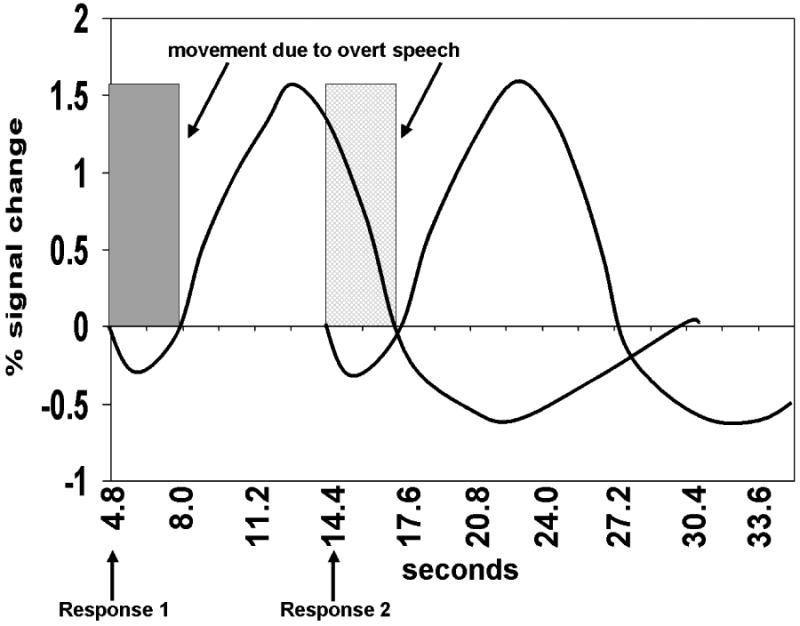
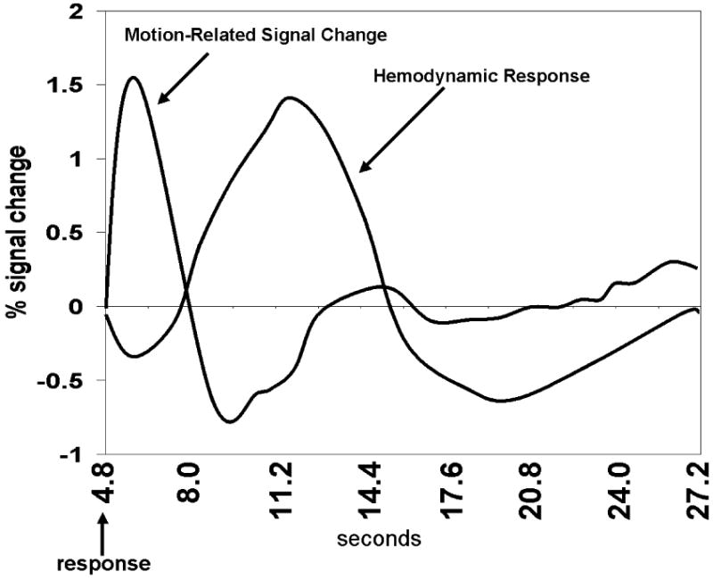
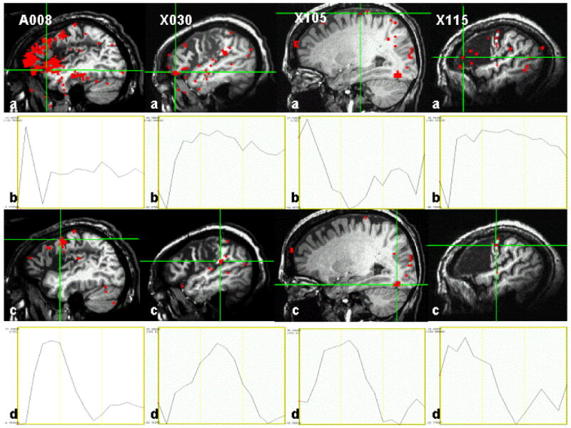
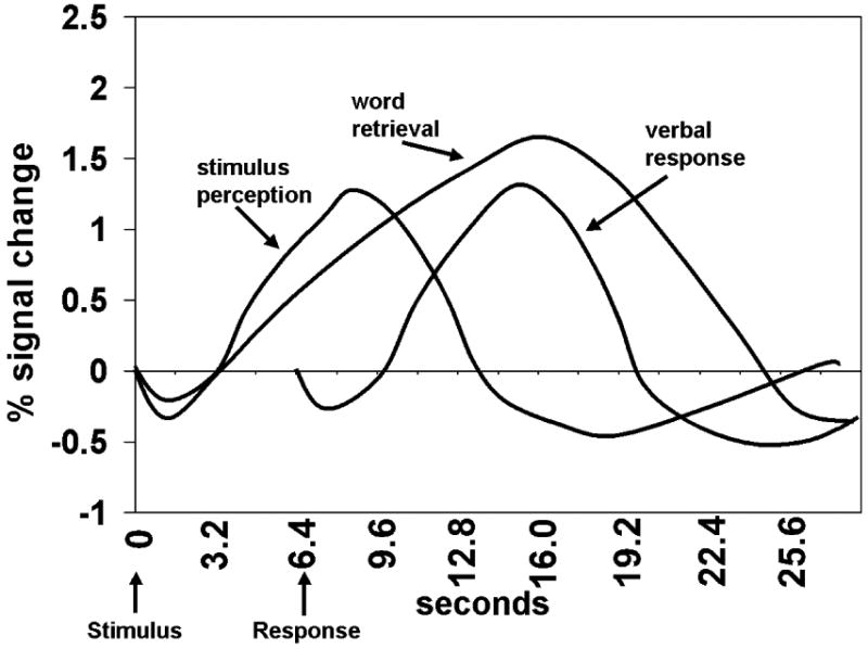
Animal analogue studies show that damaged adult brains reorganize to accommodate compromised functions. In the human arena, functional magnetic resonance imaging (fMRI) and other functional neuroimaging techniques have been used to study reorganization of language substrates in aphasia. The resulting controversy regarding whether the right or the left hemisphere supports language recovery and treatment progress must be reframed. A more appropriate question is when left-hemisphere mechanisms and when right-hemisphere mechanisms support recovery of language functions. Small lesions generally lead to good recoveries supported by left-hemisphere mechanisms. However, when too much language eloquent cortex is damaged, right-hemisphere structures may provide the better substrate for recovery of language. Some studies suggest that recovery is particularly supported by homologues of damaged left-hemisphere structures. Evidence also suggests that under some circumstances, activity in both the left and right hemispheres can interfere with recovery of function. Further research will be needed to address these issues. However, daunting methodological problems must be managed to maximize the yield of future fMRI research in aphasia, especially in the area of language production. In this review, we cover six challenges for imaging language functions in aphasia with fMRI, with an emphasis on language production: (1) selection of a baseline task, (2) structure of language production trials, (3) mitigation of motion-related artifacts, (4) the use of stimulus onset versus response onset in fMRI analyses, (5) use of trials with correct responses and errors in analyses, and (6) reliability and stability of fMRI images across sessions. However, this list of methodological challenges is not exhaustive. Once methodology is advanced, knowledge from conceptually driven fMRI studies can be used to develop theoretically driven, mechanism-based treatments that will result in more effective therapy and to identify the best patient candidates for specific treatments. While the promise of fMRI in the study of aphasia is great, there is much work to be done before this technique will be a useful clinical tool.


Figures

Figure 1
Figure 1
Representation of time courses for BOLD contrast signal changes related to spoken production of words in successive trials are shown. Times of response initiation are indicated by arrows below the time axis of the plot in seconds after stimulus presentation. The shaded area represents the region of the first time course vulnerable to motion-related signal artifacts from overt language production. If a second trial is presented too rapidly after the first trial, the latter portion of the HDR for the first trial may become contaminated by motion-related signal changes from the second trial. The cross-hatched region represents the portion of the time course vulnerable to motion-related signal changes from a second trial presented too rapidly after the first.
Figure 2
Figure 2
Representation of time courses of a motion-related signal change and of a true BOLD hemodynamic response in an area of brain participating in production of a verbal response. Time from presentation of the stimulus is shown on the horizontal axis. The more rapid evolution of the motion-related signal change than of the BOLD response can be used to reduce motion-related artifacts by various analysis methods.
Figure 3
Figure 3
Examples of motion-related signal change and HDRs for each participant (response-locked analyses). The four columns each represent a different aphasic patient. The first (top) row (a) shows images of significant signal change before selective detrending was applied to the data. The second row (b) shows the deconvolved time course of the voxel at the cross hairs in the image just above it (a). The third row (c) shows images of significant signal change after selective detrending has been applied to the data to remove motion-related signal changes. Note that the detrended images have lost many voxels of significant signal change that represent motion-related signal change rather than HDRs. Many voxels eliminated by selective detrending were in areas of lesion or were outside the brain. The fourth (bottom) row (d) shows the deconvolved time course of the voxel at the cross hairs in the image just above it (c). Note that for motion-related signal, change is most dramatic in the first 3 images after the spoken response; however, hemodynamic responses have a characteristically extended time course across several images. Thresholds for significant activity (red) were set at R2=.20 for word generation and R2=.16 for picture naming because of the differences in sensitivity between paradigms.
Figure 4
Figure 4
Representation of time courses of BOLD contrast signal changes related to three different cognitive activities during a word generation trial: perceiving and comprehending the stimulus, retrieving the appropriate word for the picture or category member, and speaking (producing) the selected word. Time of stimulus presentation and response initiation are indicated by arrows below the time axis of the plot. Because of variable response latencies in aphasic patients, the onset of the response cannot be accurately predicted from stimulus onset. The BOLD response related to perceiving and comprehending the stimulus begins soon after stimulus presentation and may return to baseline independent of when the response is given. The BOLD response related to word retrieval also may begin soon after stimulus presentation; however, because cognitive processes related to word retrieval may continue until a response is given, the hemodynamic response is extended in time and may return to its baseline only after the response is given. The BOLD response related to producing the selected word begins just prior to the time of the response. The model as depicted in this figure assumes that the major difficulty for word production lies in word retrieval. Difficulties in comprehension or motor programming of a response may lengthen the hemodynamic responses for stimulus perception or verbal response, respectively.
Figure 5
Figure 5
Total volume of significant activity in selected regions of interest for response-locked (dark gray) and stimulus locked (light gray) analyses. Both A008 and X030 participated in the word-generation paradigm, where the statistical threshold was R2 ≥ .20. For subject A008, the response-locked deconvolution is clearly more sensitive than the stimulus-locked deconvolution in that 15 of the 16 active ROIs show greater activity with the response-locked than with the stimulus-locked analysis. For X030, neither analysis is clearly more sensitive. For both X105 and X115, who received the picture-naming paradigm (with a statistical threshold of R2 ≥ .16), the stimulus-locked analysis was more sensitive. For future research, these profiles suggest that both paradigmatic differences and patient variables should be explored to assess which analysis provides superior sensitivity. Also, some areas may show activity with one type of analysis but not the other; thus, the purpose of the analysis also should be considered. L = left, R = right. LF = lateral frontal, MF = medial frontal, BG = basal ganglia, Th = thalamus, PP = posterior perisylvian, OP = other parietal, Au = auditory cortex, Vi = visual cortex, Lm = perilimbic cortex.
Figure 6
Figure 6
Frontal views of pre- and post-intention treatment images for A008. Left-hemisphere activity volumes remain relatively stable from pre- to post-treatment images. However, there is a significant increase in right-hemisphere activity volumes from pre- to post-treatment images. No significant difference in lateralization of lateral frontal activity existed on the pre-treatment image; however, lateral frontal activity is significantly lateralized to the right hemisphere at post-treatment, consistent with the experimental hypothesis.

References

    1. Abo M, Senoo A, Watanabe S, Miyano S, Doseki K, Sasaki N, Kobayashi K, Kikuchi Y, Yonemoto K. Language-related brain function during word repetition in post-stroke aphasics. NeuroReport. 2004;15:1891–1894. - PubMed
    1. Barch D, Sabb F, Carter C, Braver T, Noll D, Cohen D. Overt verbal responding during fMRI scanning: Empirical investigations of problems and potential solutions. Neuroimage. 1999;10:642–657. - PubMed
    1. Barch DM, Braver TS, et al. Anterior cingulate and the monitoring of response conflict: Evidence from an fMRI study of overt verb generation. Journal of Cognitive Neuroscience. 2000;12:298–309. - PubMed
    1. Barlow T. On a case of double cerebral hemiplegia, with cerebral symmetrical lesions. British Medical Journal. 1877;2:103–104. - PMC - PubMed
    1. Basso A, Gardelli M, Grassi MP, Mariotti M. The role of the right hemisphere in recovery from aphasia. Two case studies. Cortex. 1989;25:555–566. - PubMed

Publication types

MeSH terms