. 2007 Jul 20;282(29):20868-76.
doi: 10.1074/jbc.M702547200. Epub 2007 May 25.

Functional sequestration of transcription factor activity by repetitive DNA

Affiliations

Functional sequestration of transcription factor activity by repetitive DNA

Xiaowei Liu et al. J Biol Chem. .

Abstract

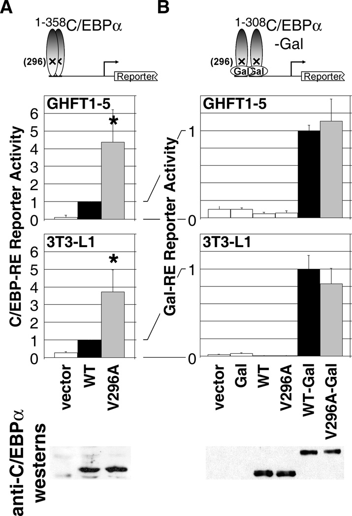
Higher eukaryote genomes contain repetitive DNAs, often concentrated in transcriptionally inactive heterochromatin. Although repetitive DNAs are not typically considered as regulatory elements that directly affect transcription, they can contain binding sites for some transcription factors. Here, we demonstrate that binding of the transcription factor CCAAT/enhancer-binding protein alpha (C/EBPalpha) to the mouse major alpha-satellite repetitive DNA sequesters C/EBPalpha in the transcriptionally inert pericentromeric heterochromatin. We find that this sequestration reduces the transcriptional capacity of C/EBPalpha. Functional sequestration of C/EBPalpha was demonstrated by experimentally reducing C/EBPalpha binding to the major alpha-satellite DNA, which elevated the concentration of C/EBPalpha in the non-heterochromatic subcompartment of the cell nucleus. The reduction in C/EBPalpha binding to alpha-satellite DNA was induced by the co-expression of the transcription factor Pit-1, which removes C/EBPalpha from the heterochromatic compartment, and by the introduction of an altered-specificity mutation into C/EBPalpha that reduces binding to alpha-satellite DNA but permits normal binding to sites in some gene promoters. In both cases the loss of alpha-satellite DNA binding coincided with an elevation in the binding of C/EBPalpha to a promoter and an increased transcriptional output from that promoter. Thus, the binding of C/EBPalpha to this highly repetitive DNA reduced the amount of C/EBPalpha available for binding to and regulation of this promoter. The functional sequestration of some transcription factors through binding to repetitive DNAs may represent an underappreciated mechanism controlling transcription output.


Figures

FIGURE 1
FIGURE 1
C/EBPα concentrates at pericentromeric heterochromatin. A, B, Wild-type and C, D, mutant C/EBPα were transiently expressed in mouse 3T3-L1 cells (A, C) or pituitary progenitor GHFT1-5 cells (B, D) at levels similar to that found following differentiation of the 3T3-L1 cells to adipocytes. Wild-type C/EBPα, detected by anti-C/EBPα antibody staining, concentrates in defined subregions of the cell nucleus that coincide with pericentromeric heterochromatin stained by the blue fluorescent DNA binding dye Hoechst 33342. C/EBPα containing valine 296 mutated to alanine (V296A) does not concentrate at pericentromeric heterochromatin. The cells and images in this figure were stained in parallel and captured/displayed under identical size and image processing conditions.
FIGURE 2
FIGURE 2
Mutations that disrupt C/EBPα concentration at pericentromeric heterochromatin also disrupt binding to α-satellite DNA. A, Diagram of the primary structure of C/EBPα and the position of point mutations used in this study relative to landmarks. TA domains, transcriptional activation domains; b, basic region; ZIP, leucine zipper. Blocks of amino acids conserved amongst C/EBP proteins from different species and with selected bZIP proteins are indicated as a black bar. Numbers represent amino acid positions. C/EBPα mutants are named according to the amino acids that were mutated (e.g. X60-62). The known activities affected by these mutations are briefly described in the Results section. B, Co-localization of antibody-stained C/EBPα with Hoechst-33342 stained C/EBPα was quantified from multiple cells as a correlation coefficient for wild-type (WT) and mutant C/EBPα expressed in GHFT1-5 cells (mean +/− sd, n>50 cells for each). A correlation coefficient of +1.0 indicates complete co-localization; 0.0 indicates no relationship; −1.0 indicates completely opposite localization. *, mutants with correlation coefficients significantly (p<<0.001) less than for wild-type C/EBPα. There is no correlation coefficient for cells transfected with vector alone since there is no C/EBPα present. C, Expression level of the same mutants monitored by western blots of nuclear extracts from transfected GHFT1-5 cells (mean quantified by densitometry from three independent experiments). D, Representative gel mobility shift of radiolabelled α-satellite DNA by wild-type C/EBPα and the indicated mutants present in the nuclear extracts of GHFT1-5 cells. The complexes formed with wild-type C/EBPα were supershifted by an antibody against C/EBPα (not shown).
FIGURE 3
FIGURE 3
Correlation of pericentromeric targeting with specific binding to α-satellite DNA. A, The V296A mutation that disrupts α-satellite DNA binding does not affect binding to some other response elements. Representative electrophoretic mobility shift (black arrow) of a radiolabelled C/EBP response element from the growth hormone (GH) promoter complexed with subsaturating amounts of extracts (0.0, 1.0, 2.0 and 4.0 µg) of transfected GHFT1-5 cells expressing equivalent levels of wild-type and V296A C/EBPα. Total extract amount was held constant with the addition of 4.0, 3.0, 2.0 or 0.0 µg of extract from sham-transfected cells. The complexes detected were supershifted with an antibody against C/EBPα (open arrow). The wild-type and V296A gel shifts shown are from the same X-ray film with an intervening series of lanes digitally removed. B, Quantitative summary of electrophoretic mobility shift assays evaluating wild-type and V296A mutant binding to the GH and PU.1 response elements (mean +/− sd of three independent experiments). No statistically significant difference (p>0.10 for all concentrations) was observed in the equilibrium binding of wild-type and V296A mutant C/EBPα to either response element. C, The addition of Pit-1, which also inhibits C/EBPα localization to pericentromeric heterochromatin (35), also blocks in vitro binding of C/EBPα to α-satellite DNA. Binding of rabbit reticulocyte-expressed C/EBPα to α-satellite DNA was inhibited in a dose dependent fashion by the addition of reticulocyte-expressed Pit-1. Total reticulocyte lysate amount was held constant by the addition of sham-translated extracts.
FIGURE 4
FIGURE 4
The euchromatically-distributed V296A mutant superactivates a promoter to which V296A and WT C/EBPα bind equivalently. A, Effect of WT or mutant C/EBPα expression in GHFT1-5 cells on chloramphenicol acetyltransferase activity expressed under the control of a single growth hormone C/EBP response element (C/EBP-RE) appended to a TATA box. Superactivation of the C/EBP-RE promoter/reporter by V296A is not detected with the V314P/L315P, L324P and L331P C/EBPα control mutants that, like V296A, are not sequestered in pericentromeric heterochromatin. Data represent the mean +/− sd from four independent experiments. B, Electrophoretic mobility shift assays show that, unlike the V296A mutant, the control mutants are not capable of binding the GH response element in the C/EBP-RE reporter. Additional lanes between the WT and V296A lanes were digitally deleted for this presentation.
FIGURE 5
FIGURE 5
Superactivity by the V296A mutation is not associated with enhanced intrinsic transcriptional activity. A, Full-length V296A superactivates the C/EBP-RE reporter (see Fig. 4) in both undifferentiated 3T3-L1 cells and GHFT1-5 cells. *, promoter activation of V296A C/EBPα that is significantly greater than WT C/EBPα. B, Intrinsic transcriptional activity was measured as the amount of luciferase expressed from a promoter containing Gal4 binding sites upon co-expression of fusions with the Gal4 DNA binding domain with amino acids 1–308 of the wild-type or V296A mutant C/EBPα. There was no activation by the Gal4 DBD alone or by either wild-type or mutant C/EBPα (amino acids 1–308). Data represent the mean +/− sd from six, (A, GHFT1-5), four (A, 3T3-L1) or three (B, GHFT1-5 and 3T3-L1) independent experiments. Representative western blots show equivalent amount of expression of the wild-type and V296A mutant C/EBPα proteins.
FIGURE 6
FIGURE 6
Reduced binding of the V296A mutant of C/EBPα to α-satellite DNA is accompanied by increased binding to and activation of the PU.1 gene. Representative chromatin immunoprecipitation of A, α-satellite and B, PU.1 promoter DNA by anti-C/EBPα antibodies in GHFT1-5 cells transfected with empty, wild-type or V296A mutant C/EBPα expression vectors. Input, 2.5% of input DNA. C, Quantification of the amounts of α-satellite DNA or PU.1 promoter precipitated (mean +/− sd from four independent experiments). Inset, representative western blot of nuclear extracts probed with an anti-C/EBPα antibody. Lane order in the inset is identical to that presented for the ChIP: vector, C/EBP, V296A. D, Quantitative RT-PCR measurement of PU.1 mRNA amount in GHFT1-5 cells transfected with an IRES-GFP expression vector, or the same vector containing wild-type or mutant C/EBPα inserted upstream of the internal ribosomal entry site. GFP fluorescence was used to sort the transfected cells away from the untransfected cells before RNA collection.
FIGURE 7
FIGURE 7
Pit-1 co-expression reduces C/EBPα binding to α-satellite DNA and increases binding to and activation of the PU.1 promoter. Representative chromatin immunoprecipitation of A, α-satellite and B, PU.1 promoter DNA by anti-C/EBPα antibodies in GHFT1-5 cells transfected with empty vector or vector expressing wild-type C/EBPα, Pit-1 or C/EBPα and Pit-1 together. Input, 2.5% of input DNA. C, Quantification of the amounts of α-satellite DNA or PU.1 promoter precipitated (mean +/− sd from three independent experiments). Inset, representative western blot of nuclear extracts probed with an anti-FLAG antibody that detects both the FLAG-tagged C/EBPα and FLAG-tagged Pit-1. Lane order in the inset is identical to that presented for the ChIP. D, Amounts of luciferase expressed from the −85/+152 PU.1 promoter in extracts of GHFT1-5 cells two days after transfection with empty vector, wild-type or V296A mutant C/EBPα, with or without co-expressed Pit-1 (mean +/− sd from six independent experiments).

Similar articles

Cited by

References

    1. Isogai Y, Tjian R. Curr Opin Cell Biol. 2003;15:1–8. - PubMed
    1. Kosak ST, Groudine M. Genes Dev. 2004;18:1371–1384. - PubMed
    1. Misteli T. Cell. 2004;119:153–156. - PubMed
    1. Henikoff S, Ahmad K. Ann Rev Cell Dev Biol. 2005;21:133–153. - PubMed
    1. Henikoff S, Ahmad K, Malik HS. Science. 2001;293:1098–1102. - PubMed

Publication types