. 2007;35(11):3859-67.
doi: 10.1093/nar/gkm337. Epub 2007 May 25.

Cell cycle regulation as a mechanism for functional separation of the apparently redundant uracil DNA glycosylases TDG and UNG2

Affiliations

Cell cycle regulation as a mechanism for functional separation of the apparently redundant uracil DNA glycosylases TDG and UNG2

Ulrike Hardeland et al. Nucleic Acids Res. 2007.

Abstract

Human Thymine-DNA Glycosylase (TDG) is a member of the uracil DNA glycosylase (UDG) superfamily. It excises uracil, thymine and a number of chemical base lesions when mispaired with guanine in double-stranded DNA. These activities are not unique to TDG; at least three additional proteins with similar enzymatic properties are present in mammalian cells. The successful co-evolution of these enzymes implies the existence of non-redundant biological functions that must be coordinated. Here, we report cell cycle regulation as a mechanism for the functional separation of apparently redundant DNA glycosylases. We show that cells entering S-phase eliminate TDG through the ubiquitin-proteasome system and then maintain a TDG-free condition until G2. Incomplete degradation of ectopically expressed TDG impedes S-phase progression and cell proliferation. The mode of cell cycle regulation of TDG is strictly inverse to that of UNG2, which peaks in and throughout S-phase and then declines to undetectable levels until it appears again just before the next S-phase. Thus, TDG- and UNG2-dependent base excision repair alternates throughout the cell cycle, and the ubiquitin-proteasome pathway constitutes the underlying regulatory system.


Figures

Figure 1.
Figure 1.
293T cells expressing high levels of TDG accumulate in S-phase. (A) 293T cells were transiently co-transfected with a plasmid overexpressing either active HA-TDG (pTDG), the catalytically inactive variant HA-TDG/N140A (pTDGN140A), or a vector control (pHH), and a EGFP expressing plasmid at a 10:1 ratio. The histogram shows the cell cycle distribution of transfected cells gated for EGFP positive cells, as determined by flow cytometry. The bottom panel documents TDG protein levels of the respective total cell population as determined by western blotting. TDG levels in cell populations carrying the overexpression construct were elevated by 20–30-fold. High levels of HA-TDG expression significantly increased the fraction of S-phase cells. This change in cell cycle distribution required TDG to be active, as overexpression of HA-TDG/N140A failed to produce the same effect. (B) The histogram shows the cell cycle distribution of 293T cells expressing active HA-TDG (pTDG) two days after transfection (2d) or after two weeks of selection for stable expression (2w). A vector control was also included (pHH). TDG expression levels are documented by western blots in the bottom panel. Shortly after transfection, TDG protein levels were 20–30 times higher than normal, but dropped to about three times the amount of endogenous TDG after selection. Concomitantly, the cell cycle effect seen after transfection disappeared. P-values (asterisk) were obtained by the Fisher's exact test from contingency tables comparing the distributions of G1-, S- and G2-cells. (Open circle) Unspecific cross-reaction of the primary antibody. (Filled square) Faster migrating forms of TDG. HA-/TDG-S: SUMO-modified HA-TDG and endogenous TDG, respectively.
Figure 2.
Figure 2.
TDG is absent in S-phase arrested HeLa cells. (A) Schematic illustration of expression of cyclin E, cyclin A and cyclin B during the cell cycle. (B and C) HeLa cells expressing endogenous TDG alone or together with HA-TDG were blocked in S-phase with hydroxyurea (HU) or in G2/M with nocodazole (NO). Untreated asynchronous cells (-) and DMSO (DM) mock-treated cells were analysed in parallel. Denaturing cell extracts were examined by western blotting with antibodies against TDG or the HA-tag as indicated. Antibodies against Cyclin E and Cyclin B1 were applied to monitor the cell cycle arrest; β-tubulin staining served as a loading control. A monoclonal anti-TDG antibody (TDGmab) detected endogenous TDG in extracts of untreated, mock treated or G2/M arrested cells, but none in extracts from S-phase arrested cells (B). Ectopically expressed HA-TDG also declined in HU arrested cells, although faint TDG (TDGmab) and HA- (HAmab) -specific signals were still discernible (C). (D) Base release assays with a fluorescent-labelled synthetic 60-mer DNA duplex document a significant reduction of G•T processing activity in nuclear extracts from HU-arrested HeLa cells. The assay was done with 25 µg of nuclear extract supplemented with 2 U of UNG2 inhibitory UGI peptide. A denaturing polyarcylamide gel with the intact DNA strand migrating at the top (S) and the cleaved products occurring as a consequence of G•T processing (P) are shown. Immunoblots of the corresponding cell extracts with TDG and MBD4-specific antibodies are shown on the right. (Filled square) Faster migrating forms of TDG. HA-/TDG-S: SUMO-modified HA-TDG and endogenous TDG, respectively.
Figure 3.
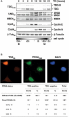
Figure 3.
Cell cycle regulation of TDG in non-arrested cells. (A) HeLa S3 cells were synchronized by mitotic shake off. Following re-plating, TDG, UNG2 and MBD4 expression was examined in a time course (TC) of 21 h. At the time point indicated, cell extracts were prepared under denaturing conditions and analysed by western blotting with specific antibodies as indicated on the left. The cell cycle phases indicated at the bottom were deduced from the expression of cyclin A (S–G2/M) and E (G1–S). β-tubulin detection served as loading control. The monoclonal anti-TDG antibody detected TDG in mitotic and G1 cells (TC 0-9) and in G2/M cells (TC 18). No TDG was detectable in S-phase cells (TC 12,15). The disappearance of TDG at 12 h coincided with the de novo expression of cyclin A, indicating a downregulation of TDG at the G1/S boundary. By contrast, nuclear UNG2 was detectable between 9 and 18 h with a peak at 12 h, representing cells in S-phase. Mitochondrial UNG1 did not fluctuate throughout the cell cycle, nor did MBD4, which shows only slightly increased expression around S-phase. (B) Immunofluorescence staining of endogenous TDG and PCNA illustrate the absence of TDG from S-phase nuclei. Upper cell, TDG positive cell with diffuse PCNA staining; lower cell, TDG negative cell with PCNA staining indicating early to mid S-phase; middle cell, TDG negative cell with fewer and larger PCNA foci indicating late S-phase. Shown are typical events of 500 randomly chosen cells scored and classified as indicated in the table at the bottom. Asterisk: statistically significant difference, P < 0.0001 by contingency tables and Fisher's exact test; TDG-S: endogenous TDG modified with SUMO.
Figure 4.
Figure 4.
TDG protein levels fluctuate during the cell cycle in primary cells but mRNA is constitutively transcribed. MRC5 primary fibroblasts were synchronized in early S-phase by serum starvation and mimosine treatment. (A) Western blot analyses of protein extracts prepared from asynchronous cells (AS), serum starved cells (ST) and cells harvested at indicated times (TP hours) after release from the mimosine block. Proteins examined were endogenous TDG, Cyclin E, Cyclin B1 and β-tubulin as a loading control. TDG-specific signals appeared at 12 h after release into S-phase and increased gradually to the levels found in the asynchronous culture. Expression of cyclin E and cyclin B1 coincided with the lack or the presence of TDG, respectively. (B) Northern blot analysis of TDG and GAPDH mRNAs (loading control) at corresponding time points, showing that TDG-specific mRNA was detectable throughout the cell cycle. TDG-S: endogenous TDG modified with SUMO.
Figure 5.
Figure 5.
TDG is polyubiquitylated and stabilized by proteasome inhibition. (A) Asynchronous HeLa cultures were treated with the proteasome inhibitor MG132 (20 µM) or DMSO. Cell extracts prepared under denaturing conditions were analysed by western blotting with a polyclonal anti-TDG (TDGab) and an anti-β-tubulin antibody (β-tubab). SUMOylated and unmodified TDG increased after proteasome inhibition. (B) HeLa cells stably transfected with a HA-TDG (pTDG) expression construct or the vector (pHH) were treated with MG132 or DMSO. Cell extracts prepared under denaturing conditions were subjected to TDG-IP with an affinity purified polyclonal anti-TDG antibody (TDGab). Bound protein fractions were analysed by western blotting with the monoclonal anti-TDG (TDGmab, left panel) or an anti-ubiquitin antibody (ubiquitinab, right panel). Strong signals appeared in the TDG-IPs but none in the IP-controls. TDG-specific signals smearing towards higher molecular weights indicated an accumulation of modified TDG in extracts of MG132 treated cells. The anti-ubiquitin antibody detected proteins with comparable migration properties in the corresponding TDG-IP protein fractions. (C) 10 ng of purified recombinant TDG were subjected to in vitro ubiquitylation. Shown is a western blot with the polyclonal anti-TDG antibody of aliquots taken at 0 and 2 h of incubation, and of a control reaction lacking TDG. The appearance of TDG-dependent high molecular weight bands after 2 h indicates ubiquitylation of TDG. (Asterisk), protein co-precipitating in TDG-IP and cross-reacting with the secondary antibody used; (Open circle) Components of the ubiquitylation system cross-reacting with the anti-TDG antibody.

Similar articles

Cited by

References

    1. Krokan HE, Drablos F, Slupphaug G. Uracil in DNA – occurrence, consequences and repair. Oncogene. 2002;21:8935–8948. - PubMed
    1. Barnes DE, Lindahl T. Repair and genetic consequences of endogenous DNA base damage in mammalian cells. Annu. Rev. Genet. 2004;38:445–476. - PubMed
    1. Slupphaug G, Eftedal I, Kavli B, Bharati S, Helle NM, Haug T, Levine DW, Krokan HE. Properties of a recombinant human uracil-DNA glycosylase from the UNG gene and evidence that UNG encodes the major uracil-DNA glycosylase. Biochemistry. 1995;34:128–138. - PubMed
    1. Haushalter KA, Todd Stukenberg MW, Kirschner MW, Verdine GL. Identification of a new uracil-DNA glycosylase family by expression cloning using synthetic inhibitors. Curr. Biol. 1999;9:174–185. - PubMed
    1. Hendrich B, Hardeland U, Ng HH, Jiricny J, Bird A. The thymine glycosylase MBD4 can bind to the product of deamination at methylated CpG sites. Nature. 1999;401:301–304. - PubMed

Publication types