

pKNOT: the protein KNOT web server
- PMID: 17526524
- PMCID: PMC1933195
- DOI: 10.1093/nar/gkm304
pKNOT: the protein KNOT web server
Abstract
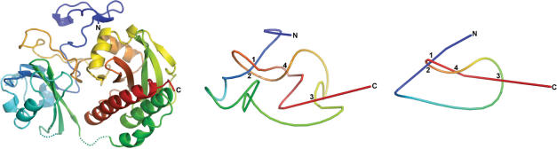
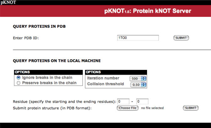
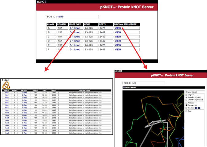
Knotted proteins are more commonly observed in recent years due to the enormously growing number of structures in the Protein Data Bank (PDB). Studies show that the knot regions contribute to both ligand binding and enzyme activity in proteins such as the chromophore-binding domain of phytochrome, ketol-acid reductoisomerase or SpoU methyltransferase. However, there are still many misidentified knots published in the literature due to the absence of a convenient web tool available to the general biologists. Here, we present the first web server to detect the knots in proteins as well as provide information on knotted proteins in PDB-the protein KNOT (pKNOT) web server. In pKNOT, users can either input PDB ID or upload protein coordinates in the PDB format. The pKNOT web server will detect the knots in the protein using the Taylor's smoothing algorithm. All the detected knots can be visually inspected using a Java-based 3D graphics viewer. We believe that the pKNOT web server will be useful to both biologists in general and structural biologists in particular.
Figures
References
-
- Ahn HJ, Eom SJ, Yoon HJ, Lee BI, Cho H, Suh SW. Crystal structure of class I acetohydroxy acid isomeroreductase from Pseudomonas aeruginosa. J. Mol. Biol. 2003;328:505–515. - PubMed
-
- Elkins PA, Watts JM, Zalacain M, van Thiel A, Vitazka PR, Redlak M, Andraos-Selim C, Rastinejad F, Holmes WM. Insights into catalysis by a knotted TrmD tRNA methyltransferase. J. Mol. Biol. 2003;333:931–949. - PubMed
-
- Lim K, Zhang H, Tempczyk A, Krajewski W, Bonander N, Toedt J, Howard A, Eisenstein E, Herzberg O. Structure of the YibK methyltransferase from Haemophilus influenzae (HI0766): a cofactor bound at a site formed by a knot. Proteins. 2003;51:56–67. - PubMed
-
- Mosbacher TG, Bechthold A, Schulz GE. Structure and function of the antibiotic resistance-mediating methyltransferase AviRb from Streptomyces viridochromogenes. J. Mol. Biol. 2005;345:535–545. - PubMed
