. 2007;35(11):3810-22.
doi: 10.1093/nar/gkm102. Epub 2007 May 25.

C-terminal regions of topoisomerase IIalpha and IIbeta determine isoform-specific functioning of the enzymes in vivo

Affiliations

C-terminal regions of topoisomerase IIalpha and IIbeta determine isoform-specific functioning of the enzymes in vivo

René M Linka et al. Nucleic Acids Res. 2007.

Abstract

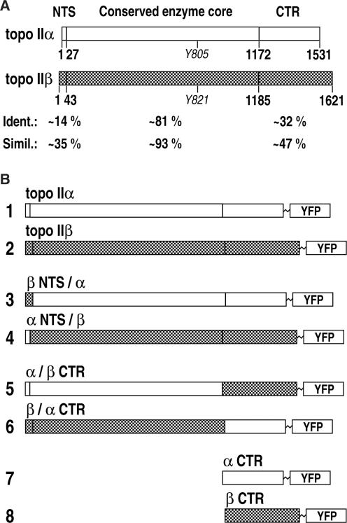
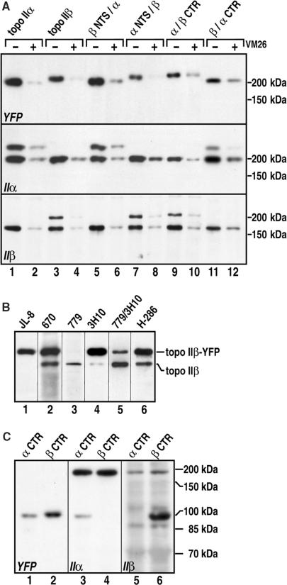
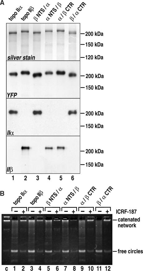
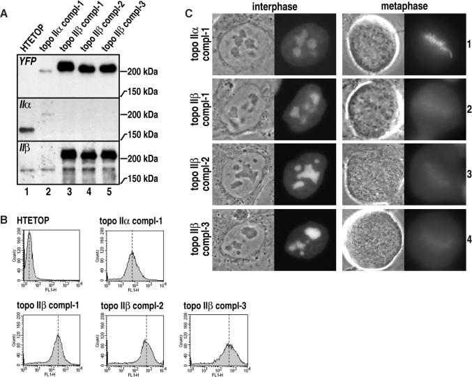
Topoisomerase II removes supercoils and catenanes generated during DNA metabolic processes such as transcription and replication. Vertebrate cells express two genetically distinct isoforms (alpha and beta) with similar structures and biochemical activities but different biological roles. Topoisomerase IIalpha is essential for cell proliferation, whereas topoisomerase IIbeta is required only for aspects of nerve growth and brain development. To identify the structural features responsible for these differences, we exchanged the divergent C-terminal regions (CTRs) of the two human isoforms (alpha 1173-1531 and beta 1186-1621) and tested the resulting hybrids for complementation of a conditional topoisomerase IIalpha knockout in human cells. Proliferation was fully supported by all enzymes bearing the alpha CTR. The alpha CTR also promoted chromosome binding of both enzyme cores, and was by itself chromosome-bound, suggesting a role in enzyme targeting during mitosis. In contrast, enzymes bearing the beta CTR supported proliferation only rarely and when expressed at unusually high levels. A similar analysis of the divergent N-terminal regions (alpha 1-27 and beta 1-43) revealed no role in isoform-specific functions. Our results show that it is the CTRs of human topoisomerase II that determine their isoform-specific functions in proliferating cells. They also indicate persistence of some functional redundancy between the two isoforms.


Figures

Figure 1.
Figure 1.
(A) Comparison of human topoisomerase IIα and IIβ. Amino acid sequence alignment of the two isoforms indicates two regions with low and one with high sequence homology. Short stretches of the N-terminal ends (denoted ‘NTS’) and the CTR have highly divergent sequences, whereas most of the ATPase domains and the catalytic cores (denoted ‘Conserved enzyme core’) are similar. Y805 and Y821 are the active site tyrosines. Numbers indicate last residues of the respective regions selected for fission/fusion and truncation of enzyme variants. Amino acid identities and similarities for each region are indicated below. (B) Schematic synopsis of the constructs studied. All constructs were fused to YFP at their C-terminal ends; topo IIα, topo IIβ: full-length human topoisomerases IIα and IIβ; β NTS/α: chimeric human topoisomerase IIβ (1–43) –> IIα (28–1531); α NTS/β: chimeric human topoisomerase IIα (1–27) −> IIβ (44–1621); α/β CTR: chimeric human topoisomerase IIα (1–1172) −> IIβ (1186–1621); β/α CTR: chimeric human topoisomerase IIβ (1–1185) −> IIα (1173–1531); α CTR: truncated human topoisomerase IIα (Δ1–1172); β CTR: truncated human topoisomerase IIβ (Δ1–1185).
Figure 2.
Figure 2.
Immunoblot analysis of YFP-fused topoisomerase II constructs expressed in HEK 293 cells. Positions of marker proteins are indicated on the right margin. See Figure 1B for nomenclature of the constructs. (A) Topoisomerase II immunoband depletion assay. Lysates of whole cells expressing full-length human topoisomerase IIα (lanes 1 and 2), or IIβ (lanes 3 and 4), or enzyme chimeras (lanes 5–12) were subjected to SDS PAGE (6% gels) and western blotting. Blots were stained with YFP antibodies (top), or antibodies against human topoisomerase IIα (middle), or IIβ (mixture of antibodies 779 and 3H10, bottom). Cells in even lanes were first cultured with 100 µM VM26 for 30 min. (B) Specificity of topoisomerase IIβ antibodies in immunostaining of western blots. Whole cell lysates of cells expressing full-length human topoisomerase IIβ, were subjected to SDS PAGE (6% gels) and western blotting. Blot membranes were probed with YFP antibodies (lane 1, JL-8) or antibodies against topoisomerase IIβ, as follows. Lane 2 (670), lane 3 (779), lane 4 (3H10), lane 5 (a mixture of antibodies 779 and 3H10) and lane 6 (H286). (C) Lysates of whole cells expressing human topoisomerase IIα Δ1–1172 (α CTR, odd numbered lanes) or IIβ Δ1–1185 (β CTR, even numbered lanes) were subjected to SDS PAGE (10% gels) and western blotting. Blots were probed with YFP-antibodies (left), or antibodies against human topoisomerase IIα (middle), or IIβ (right).
Figure 3.
Figure 3.
In vivo localization of topoisomerase II constructs in interphase nuclei and during metaphase. (A) Representative examples of HEK 293 cells stably co-expressing topoisomerase IIα fused to CFP (pseudo-colored in red) and topoisomerase IIβ fused to YFP (green) visualized by confocal imaging at interphase (left column) and mitosis (right column). (B) Each row of images shows representative images of living HEK 293 cells stably expressing the YFP-fused topoisomerase II construct indicated on the left margin (see Figure 1B for nomenclature) or YFP alone (row 9). Each pair of images visualizes the same cell by transmitted light (left) and confocal imaging of YFP-fluorescence in mid plane (right). Left and right columns of image pairs show representative examples of cells at interphase and metaphase, respectively.
Figure 4.
Figure 4.
Immunoprecipitation of YFP-fused topoisomerase II constructs followed by determination of enzyme activity in vitro. (A) Topoisomerase constructs indicated at the top (see Figure 1B for nomenclature) were stably expressed in HEK 293 cells and subjected to YFP-directed immunoprecipitation. Precipitates were analyzed by SDS PAGE (6% gels) and protein silver staining (top), or subjected to western blotting and probed with YFP antibodies (middle top), or antibodies against topoisomerase IIα (middle bottom), or IIβ (bottom). Migration distances of molecular weight marker proteins are indicated on the right margin. (B) Alternatively, precipitates were reacted with 300 ng kDNA in the absence (odd numbered lanes) or presence (even numbered lanes) of 1 mM ICRF-187. DNA-reaction products were separated by agarose gel electrophoresis and visualized with ethidium bromide. Positions of catenated DNA network and free DNA circles are indicated on the right margin. The first lane on the left (c) shows the kDNA substrate alone.
Figure 5.
Figure 5.
Characterization of HTETOP clones rescued by topoisomerase IIα or IIβ. (A) Immunoblot analysis of YFP-fused topoisomerase II constructs expressed in HTETOP cells. Whole cell lysates of untransfected HTETOP (lane 1) or tetracycline resistant HTETOP clones expressing topoisomerase IIα (lane 2) or topoisomerase IIβ (lanes 3, 4 and 5) were subjected to SDS PAGE (6% gels) and western blotting. Blots were stained with YFP antibodies (top), or antibodies against human topoisomerase IIα (middle), or IIβ (bottom). Positions of marker proteins are indicated on the right margin. (B) Yellow fluorescence level of untransfected HTETOP (HTETOP) or tetracycline resistant HTETOP clones expressing topoisomerase IIα (topo IIα compl-1) or topoisomerase IIβ (topo IIβ compl-1, topo IIβ compl-2 and topo IIβ compl-3) was measured by flow cytometry and shown as histograms. (C) In vivo localization of YFP-fused topoisomerase II constructs in interphase nuclei and during metaphase. Tetracycline resistant HTETOP clones stably expressing topoisomerase IIα (row 1) or IIβ (rows 1, 2 and 3) were visualized by fluorescence microscopy. Corresponding phase contrast images (left) and YFP images (right) are shown. Left and right of image pairs show representative examples of cells at interphase and metaphase respectively.

References

    1. Uemura T, Ohkura H, Adachi Y, Morino K, Shiozaki K, Yanagida M. DNA topoisomerase II is required for condensation and separation of mitotic chromosomes in S. pombe. Cell. 1987;50:917–925. - PubMed
    1. Holm C, Goto T, Wang JC, Botstein D. DNA topoisomerase II is required at the time of mitosis in yeast. Cell. 1985;41:553–563. - PubMed
    1. Sundin O, Varshavsky A. Arrest of segregation leads to accumulation of highly intertwined catenated dimers: dissection of the final stages of SV40 DNA replication. Cell. 1981;25:659–669. - PubMed
    1. Liu LF. DNA topoisomerase poisons as antitumor drugs. Annu. Rev. Biochem. 1989;58:351–375. - PubMed
    1. Kingma PS, Osheroff N. The response of eukaryotic topoisomerases to DNA damage. Biochim. Biophys. Acta. 1998;1400:223–232. - PubMed

Publication types

MeSH terms