

MRI of enthesitis of the appendicular skeleton in spondyloarthritis
- PMID: 17526551
- PMCID: PMC2095313
- DOI: 10.1136/ard.2007.070243
MRI of enthesitis of the appendicular skeleton in spondyloarthritis
Abstract
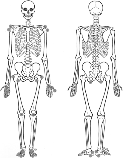
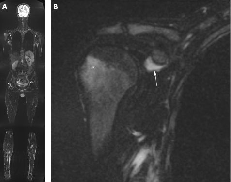
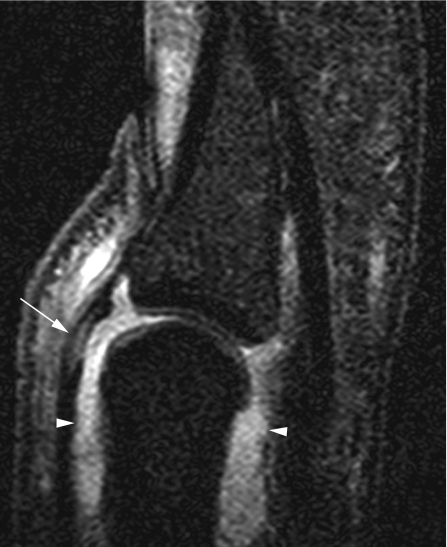
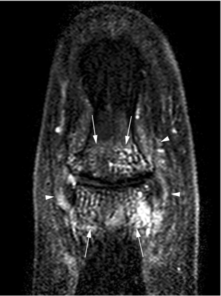
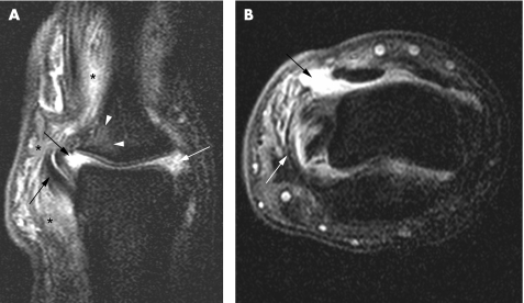
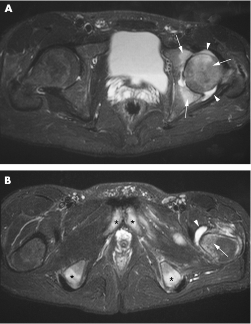
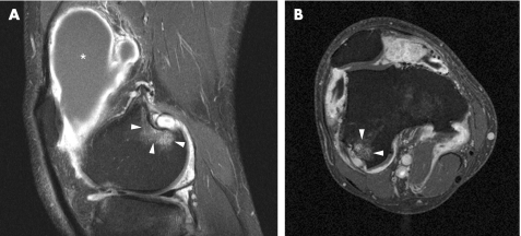
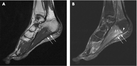
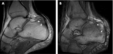
Entheses are sites where tendons, ligaments, joint capsules or fascia attach to bone. Inflammation of the entheses (enthesitis) is a well-known hallmark of spondyloarthritis (SpA). As entheses are associated with adjacent, functionally related structures, the concepts of an enthesis organ and functional entheses have been proposed. This is important in interpreting imaging findings in entheseal-related diseases. Conventional radiographs and CT are able to depict the chronic changes associated with enthesitis but are of very limited use in early disease. In contrast, MRI is sensitive for detecting early signs of enthesitis and can evaluate both soft-tissue changes and intraosseous abnormalities of active enthesitis. It is therefore useful for the early diagnosis of enthesitis-related arthropathies and monitoring therapy. Current knowledge and typical MRI features of the most commonly involved entheses of the appendicular skeleton in patients with SpA are reviewed. The MRI appearances of inflammatory and degenerative enthesopathy are described. New options for imaging enthesitis, including whole-body MRI and high-resolution microscopy MRI, are briefly discussed.
Conflict of interest statement
Competing interests: None.
References
-
- Benjamin M, Toumi H, Suzuki D, Redman S, Emery P, McGonagle D. Microdamage and altered vascularity at the enthesis‐bone interface provides an anatomic explanation for bone involvement in the HLA‐B27‐associated spondylarthritides and allied disorders. Arthritis Rheum 200756224–233. - PubMed
-
- Resnick D, Niwayama G. Entheses and enthesopathy. Anatomical, pathological, and radiological correlation. Radiology 19831461–9. - PubMed
-
- Rufai A, Ralphs J R, Benjamin M. Structure and histopathology of the insertional region of the human Achilles tendon. J Orthop Res 199513585–593. - PubMed
Publication types
MeSH terms
LinkOut - more resources
Full Text Sources
Medical
Research Materials
