. 2007 Aug 15;93(4):1264-76.
doi: 10.1529/biophysj.106.101089. Epub 2007 May 25.

Flexibility in HIV-1 assembly subunits: solution structure of the monomeric C-terminal domain of the capsid protein

Affiliations

Flexibility in HIV-1 assembly subunits: solution structure of the monomeric C-terminal domain of the capsid protein

Luis A Alcaraz et al. Biophys J. .

Abstract

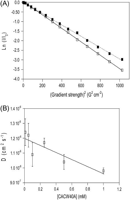
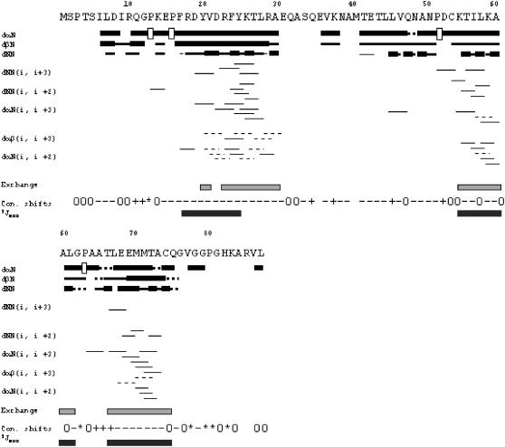
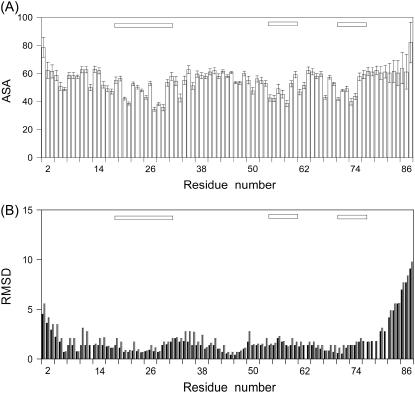
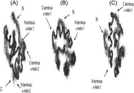
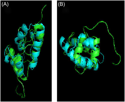
The protein CA forms the mature capsid of human immunodeficiency virus. Hexamerization of the N-terminal domain and dimerization of the C-terminal domain, CAC, occur during capsid assembly, and both domains constitute potential targets for anti-HIV inhibitors. CAC homodimerization occurs mainly through its second helix, and is abolished when its sole tryptophan is mutated to alanine. Previous thermodynamic data obtained with the dimeric and monomeric forms of CAC indicate that the structure of the mutant resembles that of a monomeric intermediate found in the folding and association reactions of CAC. We have solved the three-dimensional structure in aqueous solution of the monomeric mutant. The structure is similar to that of the subunits in the dimeric, nonmutated CAC, except the segment corresponding to the second helix, which is highly dynamic. At the end of this region, the polypeptide chain is bent to bury several hydrophobic residues and, as a consequence, the last two helices are rotated 90 degrees when compared to their position in dimeric CAC. The previously obtained thermodynamic data are consistent with the determined structure of the monomeric mutant. This extraordinary ability of CAC to change its structure may contribute to the different modes of association of CA during HIV assembly, and should be taken into account in the design of new drugs against this virus.


Figures

FIGURE 1
FIGURE 1
DOSY-NMR experiments of CACW40A. (A) The logarithm of the normalized intensity of the most upfield-shifted peaks is shown as a function of the squares of the gradient strengths at two concentrations: 1 mM (continuous line and open squares) and 500 μM (dotted line and solid squares). The slopes of the plots give the apparent diffusion constant of the molecule in solution at the particular concentration used. (B) NMR diffusion coefficients of the protein as a function of protein concentration. The bars are fitting errors to Eq. 1. The solid line is the fitting to a linear equation whose y-axis intersection yields the diffusion coefficient at 0 M protein concentration.
FIGURE 2
FIGURE 2
Summary of NMR data. NOEs are classified into strong, medium, or weak, according to the height of the of bar. The corresponding sequential Hα NOEs with the following Hδ of the proline residue are indicated by an open bar in the row corresponding to the αN (i, i+1) NOEs. The light bars indicate residues detected in a two-dimensional NOESY experiment after 4 h of exchange. Residues whose conformational shifts were >0.1, positive or negative, are represented by a + or −, respectively; 0 indicates those residues whose conformational shifts are (positive or negative) <0.1; and the * indicates the glycine residues, with two different chemical shifts for each Hα proton. The 3JNHHα are represented by a dark bar for those residues with a value <5.5 Hz, as measured from HNHA experiments.
FIGURE 3
FIGURE 3
Solvent-accessible surface area (A) and root mean-square deviations from mean coordinates (B) per residue. Calculation of the solvent-accessible surface area was carried out with MOLMOL (using a solvent radius of 1.4 Å) (48). The main-chain and side-chain RMS deviations (in Å) are indicated in black and red, respectively. The bars at the top of the panels indicate the α-helices.
FIGURE 4
FIGURE 4
Structure of CACW40A. Different views of the ensemble of the best 30 structures of CACW40A. The perpendicular arrangement of the last two helices with the first one is best shown in panel A; the parallel arrangement between the last two helices is best shown in panel B; and, the proximity between the long disordered loop and the first helix is best shown in panel C. The figures were produced with MOLMOL (48). The N and C letters indicate the N- and C-termini of the protein, respectively.
FIGURE 5
FIGURE 5
Comparison of the structures of each subunit in nonmutated, dimeric CAC and in monomeric CACW40A. (A) Ribbon diagrams of the superposition of a subunit in dimeric CAC (Protein Data Bank: 1A43) (light blue) with the best structure of CACW40A (green). (B) The same superposition of the structures in panel A, as viewed from the bottom of the orientation shown in panel A. The figures were produced with PyMOL (47), and the CAC structure was protonated with MOLMOL (48).

References

    1. Freed, E. O. 1998. HIV-1 Gag proteins: diverse functions in the virus life cycle. Virology. 251:1–15. - PubMed
    1. Freed, E. O., and M. Martin. 2001. HIVs and their replication. In Field Virology. D. M. Knipe and P. M. Howley, editors. Lippincott, Philadelphia, PA.
    1. Vogt, V. M. 1996. Proteolytic processing and particle maturation. Curr. Top. Microbiol. Immunol. 214:95–132. - PubMed
    1. Fuller, T. W., T. Wilk, B. E. Gowen, H.-G. Kräusslich, and V. M. Vogt. 1997. Cryo-electron microscopy reveals ordered domains in the immature HIV-1 particle. Curr. Biol. 7:729–738. - PubMed
    1. Ehrlich, L. S., B. E. Agresta, and C. A. Carter. 1992. Assembly of recombinant human immunodeficiency virus type 1 capsid protein in vitro. J. Virol. 66:4874–4883. - PMC - PubMed

Publication types

LinkOut - more resources