. 2007 Sep 1;93(5):1834-44.
doi: 10.1529/biophysj.107.107557. Epub 2007 May 25.

R-92L and R-92W mutations in cardiac troponin T lead to distinct energetic phenotypes in intact mouse hearts

Affiliations

R-92L and R-92W mutations in cardiac troponin T lead to distinct energetic phenotypes in intact mouse hearts

Huamei He et al. Biophys J. .

Abstract

It is now known that the flexibility of the troponin T (TnT) tail determines thin filament conformation and hence cross-bridge cycling properties, expanding the classic structural role of TnT to a dynamic role regulating sarcomere function. Here, using transgenic mice bearing R-92W and R-92L missense mutations in cardiac TnT known to alter the flexibility of the TnT tropomyosin-binding domain, we found mutation-specific differences in the cost of contraction at the whole heart level. Compared to age- and gender-matched sibling hearts, mutant hearts demonstrate greater ATP utilization measured using (31)P NMR spectroscopy as decreases in [ATP] and [PCr] and |DeltaG(~ATP)| at all workloads and profound systolic and diastolic dysfunction at all energetic states. R-92W hearts showed more severe energetic abnormalities and greater contractile dysfunction than R-92L hearts. The cost of increasing contraction was abnormally high when [Ca(2+)] was used to increase work in mutant hearts but was normalized with supply of the beta-adrenergic agonist dobutamine. These results show that R-92L and R-92W mutations in the TM-binding domain of cardiac TnT alter thin filament structure and flexibility sufficiently to cause severe defects in both whole heart energetics and contractile performance, and that the magnitude of these changes is mutation specific.


Figures

FIGURE 1
FIGURE 1
Representative 31P NMR spectra from three to six 23-week-old nTG (upper panels), R-92L (middle panels) and R-92W (lower panels) mutant hearts at baseline (left) and at high workload (right) to 4 mM Ca2+ (A) and in response to 300 nM dobutamine (B). Resonance areas from left to right correspond to Pi, PCr, and γ−, α, and β-phosphates of ATP. At baseline, R-92L hearts showed higher Pi resonance area, but lower PCr and ATP resonance areas than did nTG hearts, R-92W hearts demonstrated even greater differences compared with R-92L hearts. nTG hearts exhibited an increase in Pi resonance area, and decreases in PCr resonance area, but similar ATP resonance area at high workload in both A and B compared with at baseline. R-92L and R-92W hearts showed even greater changes in Pi, PCr and ATP resonance areas compared with at baseline as well as nTG hearts at high workload. PPM, parts per million.
FIGURE 2
FIGURE 2
Representative tracings of isovolumic contractile performance from three to six 23-week-old nTG (left column), R-92L (middle column) and R-92W (right column) mutant hearts at baseline (left) and at high workload (right) in response to 4 mM Ca2+ (A) and to 300 nM dobutamine (B). The tracings from the top to the bottom correspond to LVSP, HR, the minimum and maximum values within a beat of the first derivative of LV pressure (+dP/dt and −dP/dt). R-92L hearts showed the decreases in LVSP, +dp/dt and −dp/dt at baseline, and decreased response to 4 mM Ca2+ and to 300 nM dobutamine compared with nTG hearts. R-92W hearts demonstrated even greater decrease in LVSP, +dp/dt and −dp/dt at baseline and worse response to 4 mM Ca2+ and to 300 nM dobutamine compared toR-92L hearts. The heart was paced at 420 bpm at baseline and at high workload in response to 4 mM Ca2+. The heart was not paced when challenged with 300 nM dobutamine.
FIGURE 3
FIGURE 3
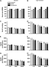
Contractile performance of hearts bearing R-92 cTnT mutations under baseline perfusion conditions (left columns) and at high workload (right columns) in response to either 4 mM Ca2+ when not paced or paced at 420 bpm or 300 nM Dobutamine. Data shown are mean ± SE, n = 3–6. For the pairs from top to bottom: (A) heart rate at baseline and (B) heart rate at high workload; (C) developed pressure (difference between left ventricular systolic pressure and end diastolic pressure) at baseline and (D) developed pressure at high workload; (E) +dp/dt at baseline, and (F) +dp/dt at high workload; (G) −dp/dt at baseline, and (H) −dp/dt at high workload. *p < 0.05; p < 0.01 vs nTG; p < 0.05; p < 0.01 vs. baseline.
FIGURE 4
FIGURE 4
Plots of the relationship between RPP and |ΔG∼ATP| for nTG, R-92L and R-92W hearts under the three protocols: unpaced +4 mM Ca2+, paced +4 mM Ca2+ and paced + 300 nM dobutamine. RPP was from supplementary Table 1, |ΔG∼ATP| was calculated from the data in Table 2. The equations, the inverse of the slopes, the assessment of goodness for the linear fits of data by group and by protocol are summarized in Table 3. (A) hearts from nTG (open circles), R-92L (solid squares), and R-92W (solid triangles) were not paced both at baseline and high workload in response to 4 mM Ca2+; (B) hearts from nTG (open circles), R-92L (solid squares) and R-92W (solid triangles) were paced both at baseline and high workload in response to 4 mM Ca2+; (C) hearts from nTG (open circles), R-92L (solid squares), and R-92W (solid triangles) were paced at baseline but not paced at high workload in response to 300 nM dobutamine; (D) nTG hearts were perfused by three protocols: unpaced-high calcium (open circles), paced-high calcium (solid squares) and dobutamine (solid triangles); (E) R-92L hearts were perfused by three protocols: unpaced-high calcium (open circles), paced-high calcium (solid squares), and dobutamine (solid triangles); (F) R-92W hearts were perfused by three protocols: unpaced-high calcium (open circles), paced-high calcium (solid squares), and dobutamine (solid triangles). The number of trials is three to six.

References

    1. Palm, T., S. Graboski, S. E. Hitchcock-DeGregori, and N. J. Greenfield. 2001. Disease-causing mutations in cardiac troponin T: identification of a critical tropomyosin-binding region. Biophys. J. 81:2827–2837. - PMC - PubMed
    1. Maytum, R., M. A. Geeves, and S. S. Lehrer. 2002. A modulatory role for the troponin T tail domain in thin filament regulation. J. Biol. Chem. 277:29774–29780. - PubMed
    1. Tobacman, L. S., M. Nihli, C. Butters, M. Heller, V. Hatch, R. Craig, W. Lehman, and E. Homsher. 2002. The troponin tail domain promotes a conformational state of the thin filament that suppresses myosin activity. J. Biol. Chem. 277:27636–27642. - PubMed
    1. Tardiff, J. C. 2005. Sarcomeric proteins and familial hypertrophic cardiomyopathy: linking mutations in structural proteins to complex cardiovascular phenotypes. Heart Fail. Rev. 10:237–248. - PubMed
    1. Javadpour, M. M., J. C. Tardiff, I. Pinz, and J. S. Ingwall. 2003. Decreased energetics in murine hearts bearing the R92Q mutation in cardiac troponin T. J. Clin. Invest. 112:768–775. - PMC - PubMed

Publication types

LinkOut - more resources