Comparative Study
. 2007 Aug;189(15):5452-62.
doi: 10.1128/JB.00408-07. Epub 2007 May 25.

Inactivation of the DnaB helicase leads to the collapse and degradation of the replication fork: a comparison to UV-induced arrest

Affiliations
Comparative Study

Inactivation of the DnaB helicase leads to the collapse and degradation of the replication fork: a comparison to UV-induced arrest

Jerilyn J Belle et al. J Bacteriol. 2007 Aug.

Abstract

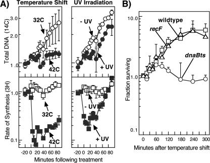
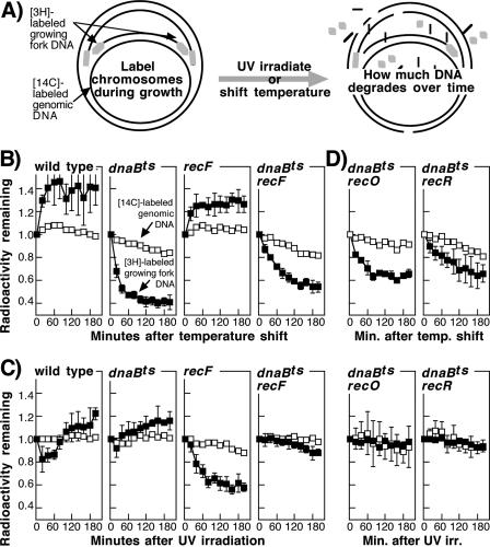
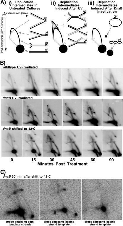
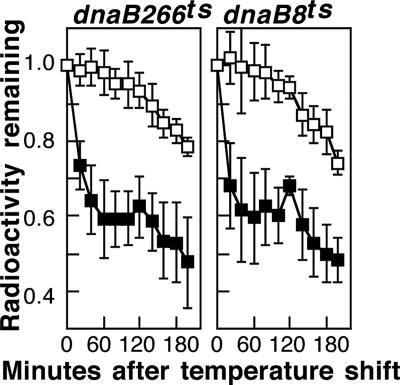
Replication forks face a variety of structurally diverse impediments that can prevent them from completing their task. The mechanism by which cells overcome these hurdles is likely to vary depending on the nature of the obstacle and the strand in which the impediment is encountered. Both UV-induced DNA damage and thermosensitive replication proteins have been used in model systems to inhibit DNA replication and characterize the mechanism by which it recovers. In this study, we examined the molecular events that occur at replication forks following inactivation of a thermosensitive DnaB helicase and found that they are distinct from those that occur following arrest at UV-induced DNA damage. Following UV-induced DNA damage, the integrity of replication forks is maintained and protected from extensive degradation by RecA, RecF, RecO, and RecR until replication can resume. By contrast, inactivation of DnaB results in extensive degradation of the nascent and leading-strand template DNA and a loss of replication fork integrity as monitored by two-dimensional agarose gel analysis. The degradation that occurs following DnaB inactivation partially depends on several genes, including recF, recO, recR, recJ, recG, and xonA. Furthermore, the thermosensitive DnaB allele prevents UV-induced DNA degradation from occurring following arrest even at the permissive temperature, suggesting a role for DnaB prior to loading of the RecFOR proteins. We discuss these observations in relation to potential models for both UV-induced and DnaB(Ts)-mediated replication inhibition.


Figures

FIG. 1.
FIG. 1.
(A) Both UV irradiation and inactivation of DnaB266 at 42°C arrest DNA synthesis. Cultures of dnaB(Ts) strain CRT266 grown at 32°C in medium containing [14C]thymine were either UV irradiated with 27 J/m2, shifted to 42°C, or mock treated. At the indicated times, duplicate aliquots of each culture were pulse-labeled for 2 min with [3H]thymidine, and the relative amounts of 14C and 3H in the DNA were determined and plotted over time. Open circles, total DNA in mock-treated cultures; filled circles, total DNA in UV-irradiated or temperature-shifted cultures; open squares, rate of DNA synthesis in mock-treated cultures; filled squares, rate of DNA synthesis in UV-irradiated or temperature-shifted cultures. The symbols indicate averages of three independent experiments. The error bars indicate one standard deviation. The 14C and 3H counts at the time of treatment ranged between 1,281 and 2,871 cpm and between 722 and 3,205 cpm, respectively, in all experiments. (B) dnaB(Ts) mutants remain viable following a temperature shift to 42°C. The fractions of cells surviving per ml of culture following incubation at 42°C for the indicated times are plotted. Circles, dnaB(Ts) strain CRT266; squares, wild-type strain SR108; triangles, recF strain HL946.
FIG. 2.
FIG. 2.
Inactivation of DnaB(Ts) at 42°C leads to extensive degradation of the nascent DNA at the arrested replication fork but prevents nascent DNA degradation from occurring after UV irradiation at the permissive temperature. (A) Schematic diagram of how the chromosome was labeled before UV irradiation or a temperature shift to 42°C. [3H]thymidine was added to [14C]thymine-prelabeled cultures for 5 s immediately before cells were filtered, resuspended in nonradioactive medium, and then either UV irradiated with 27 J/m2 or shifted to 42°C. To measure the extent of DNA degradation, the fraction of acid-precipitable radioactivity remaining in the DNA was followed over time. (B) Inactivation of DnaB(Ts) leads to extensive nascent DNA degradation following inactivation at 42°C. The relative amounts of degradation in the nascent DNA (filled symbols) and total genomic DNA (open symbols) are plotted for wild-type strain SR108, dnaB(Ts) strain CRT266, recF strain CL579, and dnaB(Ts) recF strain CL858 cultures following a temperature shift to 42°C. (C) dnaB(Ts) allele prevents the nascent DNA degradation that occurs at replication forks arrested by UV-induced damage. Degradation is plotted as described above for panel B for wild-type strain SR108, dnaB(Ts) strain CRT266, recF strain CL579, and dnaB(Ts) recF strain CL858 cultures after UV irradiation with 27 J/m2. (D) Similar to recF mutants, recO and recR mutants reduce or prevent the nascent DNA degradation from occurring at replication forks in dnaB(Ts) mutants following a temperature shift or UV irradiation. Degradation is plotted for dnaB(Ts) recO strain CL896 and dnaB(Ts) recR strain CL897 cultures after a temperature shift to 42°C or UV irradiation with 27 J/m2. The symbols indicate the averages of at least three independent experiments. The error bars indicate one standard deviation.
FIG. 3.
FIG. 3.
Exonuclease I and the RecF pathway gene products contribute to the nascent DNA degradation at replication forks disrupted following DnaB(Ts) inactivation. Degradation was measured as described in the legend to Fig. 2B. (A) Relative amounts of degradation in the nascent DNA (filled symbols) and total genomic DNA (open symbols) plotted over time for dnaB(Ts) strain CRT266, dnaB(Ts) recD strain CL743, dnaB(Ts) xonA strain CL774, dnaB(Ts) recJ strain CL742, and dnaB(Ts) xonA recJ strain CL785 following a temperature shift to 42°C. (B) Relative amounts of degradation in the nascent DNA (filled symbols) and total genomic DNA (open symbols) plotted over time for dnaB(Ts) recG strain CL1024, dnaB(Ts) ruvAB strain CL1026, and dnaB(Ts) recA strain CL1028 following a temperature shift to 42°C. The symbols indicate the averages of three independent experiments. The error bars indicate one standard deviation. The dnaB(Ts) strain CRT266 plots in panels A and B were generated by separate investigators.
FIG. 4.
FIG. 4.
Structures of plasmid replication intermediates observed following DnaB(Ts) inactivation and following UV irradiation are distinct. (A) Diagram of the migration pattern for PvuII-digested pBR322 observed by two-dimensional agarose gel electrophoresis in (i) untreated cultures, (ii) cultures following UV irradiation, and (iii) cultures following DnaB(Ts) inactivation. Nonreplicating molecules form a prominent spot that migrates as a linear 4.4-kb fragment. In untreated cultures, replicating molecules migrate more slowly due to their larger size and nonlinear shape, forming an arc that extends out from the linear fragment (approximating a simple Y arc consisting of Y-shaped molecules). Following UV irradiation, transient replication intermediates migrating in a cone-shaped region beyond the Y arc are observed at times prior to the recovery of replication and are made up of double Y- and X-shaped molecules. Following inactivation of DnaB, accumulation of an intermediate that migrates similar to circular, supercoiled plasmid molecules and that is resistant to digestion by PvuII is observed. (B) Cone region intermediates are observed following UV-induced arrest, whereas DnaB inactivation leads to a distinct circular replication intermediate that is resistant to digestion by restriction enzymes. Cultures of wild-type strain SR108 or dnaB(Ts) strain CRT266 containing plasmid pBR322 were either UV irradiated or shifted to 42°C. At the times indicated, DNA was purified, digested with PvuII, and analyzed by two-dimensional agarose gel electrophoresis using the pBR322 plasmid as a probe. (C) Circular pBR322 replication intermediate that accumulates following DnaB(Ts) inactivation is single-stranded DNA matching the lagging-strand template of the plasmid. Cultures of dnaB(Ts) strain CRT266 containing plasmid pBR322 were shifted to 42°C for 30 min before the DNA was purified and digested with PvuII. Samples were then split and analyzed by two-dimensional agarose gel electrophoresis using either the pBR322 plasmid, an oligonucleotide that is complementary to the lagging-strand template, or an oligonucleotide that is complementary to the leading-strand template as a probe.
FIG. 5.
FIG. 5.
Both dnaB266 and dnaB8 alleles result in degradation of the nascent DNA following inactivation at 42°C. Degradation was measured as described in the legend to Fig. 2B. The relative amounts of degradation in the nascent DNA (filled symbols) and total genomic DNA (open symbols) are plotted over time for dnaB266 strain CRT266 and dnaB8 strain PC8 following a temperature shift to 42°C. The symbols indicate the averages of two independent experiments. The error bars indicate one standard deviation.
FIG. 6.
FIG. 6.
Model of enzymatic activities detected at replication forks arrested by (A) UV-induced damage or (B) inactivation of DnaB(Ts). (A) (i) Following UV irradiation, the leading-strand polymerase is blocked at the site of a lesion. (ii) RecF (F), RecO (O), and RecR (R) load RecA (A) at the arrested site, limiting the nascent DNA degradation by the RecQ helicase (Q) and RecJ nuclease (J). (iii) Processing by the RecF pathway genes restores the region to a form that allows repair enzymes to remove the blocking lesion. (iv) Replication can then resume once the replication holoenzyme is reloaded. (B) (i) Inactivation of DnaB(Ts) arrests DNA synthesis. (ii) RecF, RecO, and RecR load RecA at the arrested site, limiting the nascent DNA degradation by the RecQ helicase and RecJ nuclease. (iii) Extensive degradation by exonuclease I (ExoI), a 3′-5′ exonuclease, occurs on the nascent leading-strand DNA. (iv) The amount of degradation leads to further breakdown and possible collapse of the replication fork, requiring recombination in order to restore the replication fork.

Similar articles

Cited by

References

    1. Baharoglu, Z., M. Petranovic, M. J. Flores, and B. Michel. 2006. RuvAB is essential for replication forks reversal in certain replication mutants. EMBO J. 25:596-604. - PMC - PubMed
    1. Bedale, W. A., R. B. Inman, and M. M. Cox. 1993. A reverse DNA strand exchange mediated by recA protein and exonuclease I. The generation of apparent DNA strand breaks by recA protein is explained. J. Biol. Chem. 268:15004-15016. - PubMed
    1. Bork, J. M., M. M. Cox, and R. B. Inman. 2001. The RecOR proteins modulate RecA protein function at 5′ ends of single-stranded DNA. EMBO J. 20:7313-7322. - PMC - PubMed
    1. Capaldo, F. N., and S. D. Barbour. 1975. The role of the rec genes in the viability of Escherichia coli K12. Basic Life Sci. 5A:405-418. - PubMed
    1. Carl, P. L. 1970. Escherichia coli mutants with temperature-sensitive synthesis of DNA. Mol. Gen. Genet. 109:107-122. - PubMed

Publication types

MeSH terms

LinkOut - more resources