. 2007 Aug;189(15):5716-27.
doi: 10.1128/JB.00060-07. Epub 2007 May 25.

Interactions between the lipoprotein PilP and the secretin PilQ in Neisseria meningitidis

Affiliations

Interactions between the lipoprotein PilP and the secretin PilQ in Neisseria meningitidis

Seetha V Balasingham et al. J Bacteriol. 2007 Aug.

Abstract

Neisseria meningitidis can be the causative agent of meningitis or septicemia. This bacterium expresses type IV pili, which mediate a variety of functions, including autoagglutination, twitching motility, biofilm formation, adherence, and DNA uptake during transformation. The secretin PilQ supports type IV pilus extrusion and retraction, but it also requires auxiliary proteins for its assembly and localization in the outer membrane. Here we have studied the physical properties of the lipoprotein PilP and examined its interaction with PilQ. We found that PilP was an inner membrane protein required for pilus expression and transformation, since pilP mutants were nonpiliated and noncompetent. These mutant phenotypes were restored by the expression of PilP in trans. The pilP gene is located upstream of pilQ, and analysis of their transcripts indicated that pilP and pilQ were cotranscribed. Furthermore, analysis of the level of PilQ expression in pilP mutants revealed greatly reduced amounts of PilQ only in the deletion mutant, exhibiting a polar effect on pilQ transcription. In vitro experiments using recombinant fragments of PilP and PilQ showed that the N-terminal region of PilP interacted with the middle part of the PilQ polypeptide. A three-dimensional reconstruction of the PilQ-PilP interacting complex was obtained at low resolution by transmission electron microscopy, and PilP was shown to localize around the cap region of the PilQ oligomer. These findings suggest a role for PilP in pilus biogenesis. Although PilQ does not need PilP for its stabilization or membrane localization, the specific interaction between these two proteins suggests that they might have another coordinated activity in pilus extrusion/retraction or related functions.


Figures

FIG. 1.
FIG. 1.
Gene organization and pilP constructs. (A) Chromosomal organization of the pilP locus of N. meningitidis. (B) Structural features of PilP. The lipobox and proline-rich regions are shaded in gray. Diagonal and dotted boxes indicate disordered and folded regions, respectively. (C) Schematic representation of the PilP clones used in this study. Internal deletions of the PilP protein are shaded in gray. Positions of the six-His tag are marked with H's, and the kanamycin cassette insertion in N. meningitidis M1080 pilP is also indicated. Relative molecular masses in kDa are given. na, not applicable; *, the PilPΔ1-19 product contains an N-terminal His tag with 35 amino acids derived from the vector.
FIG. 2.
FIG. 2.
PilP immunodetection. (A) Purified recombinant PilP proteins were immunodetected. Lanes: 1, full-length PilP; 2, PilPΔ1-12; 3, PilPΔ1-15; 4, PilPΔ1-16; 5, PilPΔ1-77; 6, PilPΔ61-80; 7, PilPΔ114-181. (B) Detection of M1080-PilP74-6xHis with anti-PilP antiserum K824 (lanes 1 and 2) and anti-His (lanes 3 and 4). Lanes 1 and 3, wild-type M1080; lanes 2 and 4, M1080-PilP74-6xHis. PilP protein is denoted with arrowheads I and II. Positions of the molecular mass standard proteins are indicated on the left in kDa.
FIG. 3.
FIG. 3.
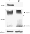
PilP is not present in OMV. Immunoblotting of the whole-cell lysates (lane 1) and purified OMV (lane 2) from N. meningitidis MC58 detected with anti-PilP antiserum (K824), anti-PilQ antiserum (K010), and anti-OpcA antibody. α, anti.
FIG. 4.
FIG. 4.
PilP copurifies with the inner membrane. The subcellular localization of N. meningitidis PilP was assessed by sucrose gradient centrifuge analysis of membrane fractions. Fractions enriched in the inner membranes were identified by monitoring LDH activity (graph, fractions 4 to 8). The presence of PilP protein was detected by immunoblotting using anti-PilP antiserum K824 (upper gel) and coincided with the LDH activity (fractions 4 to 8). Outer membrane fractions (10 to 13) enriched in the outer membrane proteins PilQ and OpcA (middle and lower gels) were detected by immunoblotting with anti-PilQ and anti-OpcA antibodies, respectively. OD/min, optical density per minute; •, LDH activity; ▪, sucrose density; α, anti.
FIG. 5.
FIG. 5.
The genes encoding PilP and PilQ are cotranscribed. RT-PCR analysis of pilP and pilQ from N. meningitidis M1080. Ten microliters of each reaction mixture was subjected to electrophoresis on a 2% agarose gel. Lanes: 1, DNA size marker; 2 and 3, RT-PCRs on RNA treated with RNase for 5 and 20 min, respectively; 4, RT-PCR on RNA template using pilP- and pilQ-specific primers pilP-5075 and pilQ-6485; 5, PCR on RNA with pilQ-specific primers pilQ-5675 and pilQ-6485 using Taq polymerase; 6, RT-PCR on RNA without pilQ reverse primer; 7, RT-PCR on RNA using pilQ-specific primers pilQ-5675 and pilQ-6485. The products in lane 6, obscuring the interpretation of results, indicate a fold-back artifact of RNA, which can provide a 3′ terminus appropriate for reverse transcriptase to act on even in the absence of reverse primer. Molecular size markers are indicated on the left in kilobases.
FIG. 6.
FIG. 6.
PilP is not required for PilQ multimer stability. Meningococcal PilQ and PilP expression in whole-cell lysates monitored by immunoblotting. (A) Samples detected with anti-PilQ antiserum (K010); (B) samples detected with anti-PilQ antiserum after phenol extraction; (C) samples detected with anti-PilP antiserum K824. Lanes: 1, wild-type M1080; 2, M1080pilP74-6xHis; 3, M1080ΔpilP; 4, M1080ΔpilP iga::pilP; 5, M1080pilPfs; 6, M1080pilPfs iga::pilP; 7, M1080ΔpilQ. Arrowheads indicate the positions of proteins. Equal amounts of starting materials were used to make cell lysates. Positions of the molecular mass standard proteins are indicated on the left in kDa.
FIG. 7.
FIG. 7.
Meningococcal PilP and PilQ monomers directly interact. Interaction of recombinant meningococcal PilP with recombinant PilQ protein detected by a solid-phase overlay assay (far-Western analysis). (A) Schematic diagram showing the pilQ gene constructs encoding the four recombinant proteins used in this study. H represents the position of the polyhistidine tag. (B) Far-Western analysis; the three panels, I to III, show gels with the same samples in each lane, but overlaid with different recombinant PilP proteins before immunodetection with anti-PilP antiserum K824. Panels: I, full-length recombinant PilP; II, recombinant PilPΔ1-12; III, recombinant PilPΔ61-80. Lanes: 1, full-length PilQ; 2, PilQ25-354; 3, PilQ217-478; 4, PilQ350-761; 5, Hemoglobin-binding outer membrane receptor protein (HmbR); 6, bovine serum albumin. Positions of the molecular mass standard proteins are indicated on the left in kDa.
FIG. 8.
FIG. 8.
3D reconstruction of the complex between PilPΔ1-19 and the PilQ oligomer. (A) Sample micrograph of PilQ particles, with PilPΔ1-19 bound, in cryonegative stain (14). The positions of individual PilQ-PilP particles are circled. Scale bar = 1,000 Å. (B) PilQ-PilP particle classification. 2D class averages for each of the particle classes following reference-free alignment are shown with the corresponding 2D back projections used for the C4 3D reconstruction. Averages were calculated by using the software package EMAN. Box size = 250 Å2. (C) Comparison of 3D reconstructions of PilQ and PilQ-PilP oligomers. Isosurface renderings of volumes are displayed at thresholds appropriate to accommodate 900 to 1,000 kDa of mass. The top structure shows the structure of the PilQ oligomer alone (14). The middle structure shows the PilQ oligomer bound to PilPΔ1-19, determined from 7,700 particles. The structure converged correctly using a symmetrical start model, a class-selected C1 start model, or an elliptical “blob”; model-dependent bias was therefore eliminated. The bottom structure shows the structure of the PilQ oligomer bound to PilPΔ1-19 and Ni-NTA-nanogold (n = 1,010). The structure was calculated in the same way from a cryonegatively stained sample containing Ni-NTA-nanogold. Scale bar = 100 Å.

References

    1. Alm, R. A., and J. S. Mattick. 1997. Genes involved in the biogenesis and function of type-4 fimbriae in Pseudomonas aeruginosa. Gene 192:89-98. - PubMed
    1. Areschoug, T., S. Linse, M. Stalhammar-Carlemalm, L.-O. Heden, and G. Lindahl. 2002. A proline-rich region with a highly periodic sequence in streptococcal β-protein adopts the polyproline II structure and is exposed on the bacterial surface. J. Bacteriol. 184:6376-6383. - PMC - PubMed
    1. Assalkhou, R., S. Balasingham, R. F. Collins, S. A. Frye, T. Davidsen, A. V. Benam, M. Bjoras, J. P. Derrick, and T. Tonjum. 2007. The outer membrane secretin PilQ from Neisseria meningitidis binds DNA. Microbiology 153:1593-1603. - PMC - PubMed
    1. Bayan, N., I. Guilvout, and A. P. Pugsley. 2006. Secretins take shape. Mol. Microbiol. 60:1-4. - PubMed
    1. Bitter, W. 2003. Secretins of Pseudomonas aeruginosa: large holes in the outer membrane. Arch. Microbiol. 179:307-314. - PubMed

Publication types

MeSH terms