. 2007 Aug;189(15):5495-503.
doi: 10.1128/JB.00190-07. Epub 2007 May 25.

The two-component regulatory system senX3-regX3 regulates phosphate-dependent gene expression in Mycobacterium smegmatis

Affiliations

The two-component regulatory system senX3-regX3 regulates phosphate-dependent gene expression in Mycobacterium smegmatis

Robert T Glover et al. J Bacteriol. 2007 Aug.

Abstract

Phosphate import is required for the growth of mycobacteria and is regulated by environmental inorganic phosphate (P(i)) concentrations, although the mechanism of this regulation has not been characterized. The expression of genes involved in P(i) acquisition is frequently regulated by two-component regulatory systems (2CRs) consisting of a sensor histidine kinase and a DNA-binding response regulator. In this work, we have identified the senX3-regX3 2CR as a P(i)-dependent regulator of genes involved in phosphate acquisition in Mycobacterium smegmatis. Characterization of senX3 mutants with different PhoA phenotypes suggests a dual role for SenX3 as a phosphatase or a phosphodonor for the response regulator RegX3, depending upon P(i) availability. Expression of PhoA activity required phosphorylation of RegX3, consistent with a role for phosphorylated RegX3 (RegX3 approximately P) as a transcriptional activator of phoA. Furthermore, purified RegX3 approximately P bound to promoter sequences from phoA, senX3, and the high-affinity phosphate transporter component pstS, demonstrating direct transcriptional control of all three genes. DNase I footprinting and primer extension analyses have further defined the DNA-binding region and transcriptional start site within the phoA promoter. A DNA motif consisting of an inverted repeat was identified in each of the promoters bound by RegX3 approximately P. Based upon our findings, we propose a model for P(i)-regulated gene expression mediated by SenX3-RegX3 in mycobacteria.


Figures

FIG. 1.
FIG. 1.
The senX3-regX3 region of M. smegmatis. senX3, regX3, and flanking genes are shown as open arrows. Vertical lines indicate sites of transposon insertion within senX3. A horizontal arrow indicates the orientation of the kanamycin-selectable marker within each transposon, with strain designations indicated above each arrow. The DNA fragments used for homologous integration (bold lines) and targeted deletions (slanted lines) are shown below the gene map.
FIG. 2.
FIG. 2.
Anaylsis of gene expression in senX3 mutant strains. (A) The PhoA-constitutive senX3::Tn5371 mutant mc25023 was transformed with a vector containing the complete senX3-regX3 operon cloned from M. smegmatis (pYUB1113) or M. tuberculosis (pYUB1114). Recombinant bacteria were grown in Pi-rich media and assayed for alkaline phosphatase activity as described in Materials and Methods. (B) Total RNA was extracted from the indicated strains, and Northern blot analysis was performed using a regX3-specific RNA probe. Ethidium bromide (EtBr) staining was used to confirm equal loading. (C) Wild-type mc2155 and the ΔsenX3 strain mc25032 were transformed with pYUB1122 (black bars) and pYUB1123 (gray bars). pYUB1122 encodes His6RegX3, while pYUB1123 encodes the phosphorylation-defective mutant His6RegX3(D52N), each under the transcriptional control of a tetracycline-inducible promoter. Protein expression was induced in the indicated cultures by the addition of 50 ng/ml anhydrotetracycline and assayed for alkaline phosphatase activity. The error bars indicate standard deviations.
FIG. 3.
FIG. 3.
Phosphate-dependent transcriptional regulation of phoA and senX3-regX3. (A) Northern blot. Total RNA was extracted from bacteria grown in medium supplemented with excess Pi (50 mM) or with no added Pi, as shown. The single-stranded RNA probes used for each blot are identified on the left. Ethidium bromide (EtBr) staining of total RNA was used to demonstrate equivalent loading of RNA. (B) Recombinant mc2155 containing the PphoA-lacZ reporter plasmid pYUB1106 was grown in the presence of excess (50 mM) Pi (gray bars) or in Pi-limiting medium (black bars). At the time points indicated, aliquots were removed and assayed for β-galactosidase activity as described in Materials and Methods. The error bars indicate standard deviations.
FIG. 4.
FIG. 4.
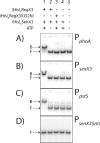
Direct binding of RegX3∼P to Pi-regulated promoter elements using EMSA. Promoter regions were amplified upstream of (A) phoA (157 bp), (B) senX3 (199 bp), and (C) pstS (139 bp) or from (D) an internal coding region of senX3 (355 bp). Radiolabeled PCR products were used in binding reactions with the protein combinations indicated above each lane. Bound (B) and Free (F) DNA is indicated on the left of each panel. Binding reactions consisted of 4 μM His6RegX3 or His6RegX3(D52N), 0.8 μM His6SenX3, and 200 pM [γ-32P]ATP-labeled DNA. The final concentration of phosphorylated His6SenX3 in lane 4 was 4 μM.
FIG. 5.
FIG. 5.
(A) DNase I footprinting of the phoA promoter bound by RegX3∼P. End-labeled phoA promoter DNA was incubated with (lane 1) 1.5 μM, (lane 2) 3 μM, (lane 3) 6 μM, (lane 4) 12 μM, or (lane 5) 24 μM phosphorylated His6RegX3. Following DNase I treatment, samples were run on a 5% acrylamide sequencing gel and visualized on a phosphorimager. The DNA region protected from DNase I cleavage is indicated. As a control, the same experiment was performed with the omission of ATP in the phosphorylation reaction. (B and C) Total RNA isolated from Pi-starved cells was used in a primer extension analysis with antisense oligonucleotide primers located within phoA (B) and senX3 (C), as indicated. Samples were run adjacent to a sequencing reaction performed using the same primer as for the reverse transcription on a 4% polyacrylamide-urea gel and visualized using a phosphorimager. The unique band identified in each primer extension is distinguished from adjacent sequence by a capital letter.
FIG. 6.
FIG. 6.
Conserved regulatory sequences identified within Pi-regulated promoters. (A) The DNA sequences used in the EMSA assay were compared using Gibbs motif analysis. Sequences were aligned based on the identified motif present in each. Nucleotides conserved between at least two of the three sequences are highlighted. The consensus motif is shown below the alignment with the inverted-repeat sequence underlined. n, unconserved nucleotide. (B) The sequence of the phoA promoter is shown with regulatory elements indicated, including the DNase I protected region, −10 consensus, TSS, RBS, and translation initiation codon.
FIG. 7.
FIG. 7.
Model of phoA regulation by SenX3-RegX3. When environmental Pi is abundant, SenX3 is required to maintain RegX3 in an unphosphorylated state, where it is unable to bind target DNA and gene transcription is repressed (1). When Pi becomes a limiting nutrient, SenX3 autophosphorylates and becomes a phosphodonor for RegX3 (2). The increased affinity of RegX3∼P for its consensus motif permits DNA binding and activates gene transcription. In the absence of functional SenX3, RegX3 can acquire phosphate from another, undetermined donor molecule (3). Because the extracellular sequence of SenX3 is only a few amino acids in length, the concentration of available Pi is probably not sensed by SenX3 directly. Instead, the Pst transporter is likely to relay this information to SenX3 and to regulate its phosphatase/phosphodonor activity toward RegX3, as indicated by arrows on either side of the Pst transporter.

Similar articles

Cited by

References

    1. Amemura, M., K. Makino, H. Shinagawa, and A. Nakata. 1990. Cross talk to the phosphate regulon of Escherichia coli by PhoM protein: PhoM is a histidine protein kinase and catalyzes phosphorylation of PhoB and PhoM open reading frame 2. J. Bacteriol. 172:6300-6307. - PMC - PubMed
    1. Andersen, A. B., L. Ljungqvist, and M. Olsen. 1990. Evidence that protein antigen b of Mycobacterium tuberculosis is involved in phosphate metabolism. J. Gen. Microbiol. 136:477-480. - PubMed
    1. Bardarov, S., J. Kriakov, C. Carriere, S. Yu, C. Vaamonde, R. A. McAdam, B. R. Bloom, G. F. Hatfull, and W. R. Jacobs, Jr. 1997. Conditionally replicating mycobacteriophages: a system for transposon delivery to Mycobacterium tuberculosis. Proc. Natl. Acad. Sci. USA 94:10961-10966. - PMC - PubMed
    1. Barletta, R. G., D. D. Kim, S. B. Snapper, B. R. Bloom, and W. R. Jacobs, Jr. 1992. Identification of expression signals of the mycobacteriophages Bxb1, L1 and TM4 using the Escherichia-Mycobacterium shuttle plasmids pYUB75 and pYUB76 designed to create translational fusions to the lacZ gene. J. Gen. Microbiol. 138:23-30. - PubMed
    1. Braibant, M., and J. Content. 2001. The cell surface associated phosphatase activity of Mycobacterium bovis BCG is not regulated by environmental inorganic phosphate. FEMS Microbiol. Lett. 195:121-126. - PubMed

Publication types

MeSH terms