

FepA- and TonB-dependent bacteriophage H8: receptor binding and genomic sequence
- PMID: 17526714
- PMCID: PMC1951831
- DOI: 10.1128/JB.00437-07
FepA- and TonB-dependent bacteriophage H8: receptor binding and genomic sequence
Abstract
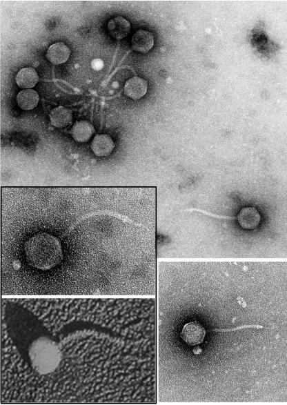
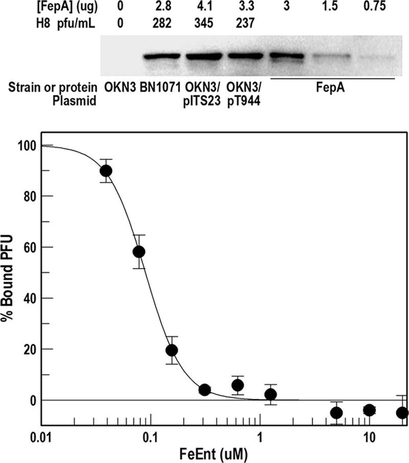
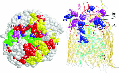
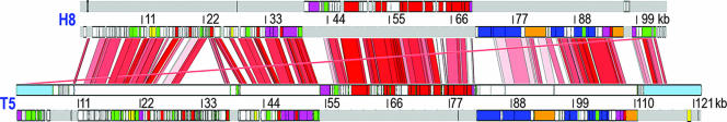
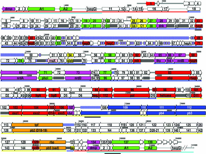
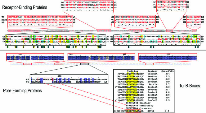
H8 is derived from a collection of Salmonella enterica serotype Enteritidis bacteriophage. Its morphology and genomic structure closely resemble those of bacteriophage T5 in the family Siphoviridae. H8 infected S. enterica serotypes Enteritidis and Typhimurium and Escherichia coli by initial adsorption to the outer membrane protein FepA. Ferric enterobactin inhibited H8 binding to E. coli FepA (50% inhibition concentration, 98 nM), and other ferric catecholate receptors (Fiu, Cir, and IroN) did not participate in phage adsorption. H8 infection was TonB dependent, but exbB mutations in Salmonella or E. coli did not prevent infection; only exbB tolQ or exbB tolR double mutants were resistant to H8. Experiments with deletion and substitution mutants showed that the receptor-phage interaction first involves residues distributed over the protein's outer surface and then narrows to the same charged (R316) or aromatic (Y260) residues that participate in the binding and transport of ferric enterobactin and colicins B and D. These data rationalize the multifunctionality of FepA: toxic ligands like bacteriocins and phage penetrate the outer membrane by parasitizing residues in FepA that are adapted to the transport of the natural ligand, ferric enterobactin. DNA sequence determinations revealed the complete H8 genome of 104.4 kb. A total of 120 of its 143 predicted open reading frames (ORFS) were homologous to ORFS in T5, at a level of 84% identity and 89% similarity. As in T5, the H8 structural genes clustered on the chromosome according to their function in the phage life cycle. The T5 genome contains a large section of DNA that can be deleted and that is absent in H8: compared to T5, H8 contains a 9,000-bp deletion in the early region of its chromosome, and nine potentially unique gene products. Sequence analyses of the tail proteins of phages in the same family showed that relative to pb5 (Oad) of T5 and Hrs of BF23, the FepA-binding protein (Rbp) of H8 contains unique acidic and aromatic residues. These side chains may promote binding to basic and aromatic residues in FepA that normally function in the adsorption of ferric enterobactin. Furthermore, a predicted H8 tail protein showed extensive identity and similarity to pb2 of T5, suggesting that it also functions in pore formation through the cell envelope. The variable region of this protein contains a potential TonB box, intimating that it participates in the TonB-dependent stage of the phage infection process.
Figures
Similar articles
-
Aromatic components of two ferric enterobactin binding sites in Escherichia coli FepA.Mol Microbiol. 2000 Sep;37(6):1306-17. doi: 10.1046/j.1365-2958.2000.02093.x. Mol Microbiol. 2000. PMID: 10998164
-
Double mutagenesis of a positive charge cluster in the ligand-binding site of the ferric enterobactin receptor, FepA.Proc Natl Acad Sci U S A. 1997 Apr 29;94(9):4560-5. doi: 10.1073/pnas.94.9.4560. Proc Natl Acad Sci U S A. 1997. PMID: 9114029 Free PMC article.
-
Identification of host receptor and receptor-binding module of a newly sequenced T5-like phage EPS7.FEMS Microbiol Lett. 2008 Dec;289(2):202-9. doi: 10.1111/j.1574-6968.2008.01397.x. FEMS Microbiol Lett. 2008. PMID: 19025561 Free PMC article.
-
Effect of loop deletions on the binding and transport of ferric enterobactin by FepA.Mol Microbiol. 1999 Jun;32(6):1153-65. doi: 10.1046/j.1365-2958.1999.01424.x. Mol Microbiol. 1999. PMID: 10383757
-
Three paradoxes of ferric enterobactin uptake.Front Biosci. 2003 Sep 1;8:s1422-36. doi: 10.2741/1233. Front Biosci. 2003. PMID: 12957833 Review.
Cited by
-
Insight from TonB hybrid proteins into the mechanism of iron transport through the outer membrane.J Bacteriol. 2008 Jun;190(11):4001-16. doi: 10.1128/JB.00135-08. Epub 2008 Apr 4. J Bacteriol. 2008. PMID: 18390658 Free PMC article.
-
Outer Membrane Porin F in E. coli Is Critical for Effective Predation by Bdellovibrio.Microbiol Spectr. 2022 Dec 21;10(6):e0309422. doi: 10.1128/spectrum.03094-22. Epub 2022 Nov 29. Microbiol Spectr. 2022. PMID: 36445149 Free PMC article.
-
Effect of a bacteriophage T5virus on growth of Shiga toxigenic Escherichia coli and Salmonella strains in individual and mixed cultures.Virol J. 2020 Jan 7;17(1):3. doi: 10.1186/s12985-019-1269-7. Virol J. 2020. PMID: 31910855 Free PMC article.
-
Mutations in the ExbB cytoplasmic carboxy terminus prevent energy-dependent interaction between the TonB and ExbD periplasmic domains.J Bacteriol. 2011 Oct;193(20):5649-57. doi: 10.1128/JB.05674-11. Epub 2011 Aug 12. J Bacteriol. 2011. PMID: 21840979 Free PMC article.
-
O antigen is the receptor of Vibrio cholerae serogroup O1 El Tor typing phage VP4.J Bacteriol. 2013 Feb;195(4):798-806. doi: 10.1128/JB.01770-12. Epub 2012 Dec 7. J Bacteriol. 2013. PMID: 23222721 Free PMC article.
References
-
- Adams, M. H. 1959. Bacteriophages. Interscience Publishers, New York, NY.
-
- Altschul, S. F., W. Gish, W. Miller, E. W. Myers, and D. J. Lipman. 1990. Basic local alignment search tool. J. Mol. Biol. 215:403-410. - PubMed
-
- Ames, G. F. 1974. Resolution of bacterial proteins by polyacrylamide gel electrophoresis on slabs. Membrane, soluble, and periplasmic fractions. J. Biol. Chem. 249:634-644. - PubMed
-
- Armstrong, S. K., C. L. Francis, and M. A. McIntosh. 1990. Molecular analysis of the Escherichia coli ferric enterobactin receptor FepA. J. Biol. Chem. 265:14536-14543. - PubMed
Publication types
MeSH terms
Substances
Associated data
- Actions
Grants and funding
LinkOut - more resources
Full Text Sources
Other Literature Sources
Molecular Biology Databases
Research Materials
