. 2007 Aug;27(15):5486-98.
doi: 10.1128/MCB.00692-07. Epub 2007 May 25.

A double-bromodomain protein, FSH-S, activates the homeotic gene ultrabithorax through a critical promoter-proximal region

Affiliations

A double-bromodomain protein, FSH-S, activates the homeotic gene ultrabithorax through a critical promoter-proximal region

Yuh-Long Chang et al. Mol Cell Biol. 2007 Aug.

Abstract

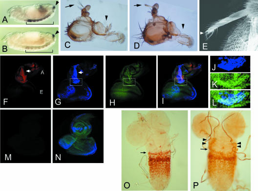
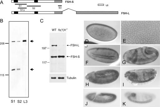
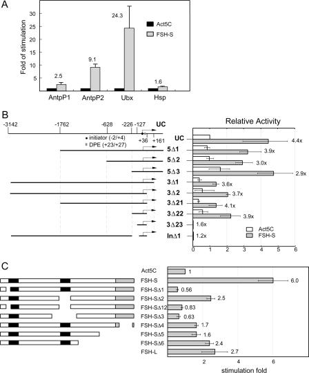
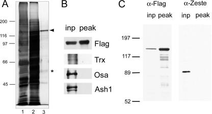
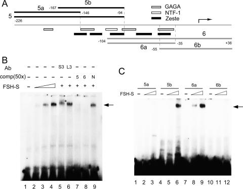
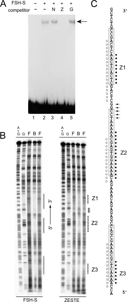
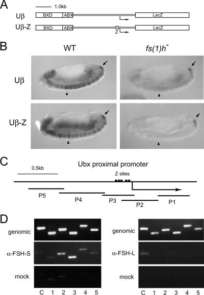
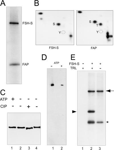
More than a dozen trithorax group (trxG) proteins are involved in activation of Drosophila HOX genes. How they act coordinately to integrate signals from distantly located enhancers is not fully understood. The female sterile (1) homeotic (fs(1)h) gene is one of the trxG genes that is most critical for Ultrabithorax (Ubx) activation. We show that one of the two double-bromodomain proteins encoded by fs(1)h acts as an essential factor in the Ubx proximal promoter. First, overexpression of the small isoform FSH-S, but not the larger one, can induce ectopic expression of HOX genes and cause body malformation. Second, FSH-S can stimulate Ubx promoter in cultured cells through a critical proximal region in a bromodomain-dependent manner. Third, purified FSH-S can bind specifically to a motif within this region that was previously known as the ZESTE site. The physiological relevance of FSH-S is ascertained using transgenic embryos containing a modified Ubx proximal promoter and chromatin immunoprecipitation. In addition, we show that FSH-S is involved in phosphorylation of itself and other regulatory factors. We suggest that FSH-S acts as a critical component of a regulatory circuitry mediating long-range effects of distant enhancers.


Figures

FIG. 1.
FIG. 1.
Essential role of fs(1)h1 in HOX gene activation. (A and B) Whole-mount in situ hybridization to reveal Ubx (bracket) and cad (arrowhead) transcripts in the ventral nerve cord of stage 13 embryos. (A) A wild-type embryo showing high levels of Ubx transcripts in PS5 to PS12. (B) An fs(1)h1 embryo showing severely reduced Ubx transcripts. No difference was found for cad transcripts between these embryos. (C to E) Head defects caused by targeted expression of FSH-S driven by dpp-Gal4. A wild-type adult head showing normal aristae (arrow) and maxillary palpus (C, arrowhead). Targeted FSH-S expression resulted in aristae-to-leg transformation and loss of maxillary palpus (D). No abnormality was observed when FSH-L was induced. Anterior is to the top. Scanning electron microscopy of ectopic leg shows a claw indicated by an arrowhead (E). (F to N) ANTP expression in antennal discs with dpp-driven FSH-S (F to L) or FSH-L (M and N). The eye-antennal discs from third instar larvae were stained by anti-ANTP (red in panels F, I, and M), anti-Flag antibodies (blue in panels G, I, J, L, and N), or a DNA dye SYTOX (green in panels H, I, K, L, and N). The lower portion of the antennal disc (boxes in G to I) is shown with higher magnification to reveal the staining of FSH-S (J) and DNA (K). The merged images of multiple staining are shown (I, L, and N). Ectopic ANTP signals were seen in antennal discs with induced FSH-S but not in wild-type discs or discs with induced FSH-L. The antennal (A) and eye (E) discs are indicated. The overlap between ANTP and FSH-S signals in the antennal disc is marked by arrows (F and G). Note that FSH-S induction also caused distortion of antennal tissue. No distortion was seen by FSH-L induction. (O and P) Ectopic UBX expression by en-driven FSH-S. Ganglia from second instar larvae were stained with an anti-UBX antibody. Normal UBX expression pattern is shown with its anterior boundary in PS5 indicated (O, arrow). Induction of FSH-S by en-Gal4 resulted in ectopic UBX signals at lateral parts of more anterior tissue (P, arrowheads). en-Gal4 is expressed in a transverse row of cells in each PS.
FIG. 2.
FIG. 2.
FSH-S is a ubiquitous nuclear protein in embryos. (A) Organization of the two ORFs of the fs(1)h gene. FSH-S contains 1,110 amino acids, while FSH-L contains an additional 932 amino acids by the alternative splicing as indicated by the overhang. Splicing junctions in other regions are omitted. The double-bromodomain, the ET domain, and the unique region of FSH-L are indicated by black, gray, and hatched boxes, respectively. S1, S2, S3, and L3 correspond to regions used for immunization. A smaller region (S1Δ) lacking the first bromodomain was used for affinity purification of S1 antibody. (B) Analysis of FSH proteins. Embryonic extracts were immunoblotted with affinity-purified S1, S2, and L3 antibodies. S1 and S2 antibodies detected both a ∼220-kDa and a ∼120-kDa protein, while the L3 antibody detected only the ∼220-kDa protein. (C) Differential effect of fs(1)h17 mutation on fs(1)h products. Extracts from wild-type or fs(1)h17/Y third instar larvae were immunoblotted with affinity-purified S3 antibody. FSH-L was severely reduced in an fs(1)h17 mutant. α-Tubulin control is shown in the bottom panel. (D to I) Embryonic patterns of FSH proteins. Wild-type embryos were stained with affinity-purified S1 antibody. FSH proteins were detected ubiquitously at blastoderm stage (D and E), beginning of gastrulation (F and G), fully extended germ-band stage (H), and dorsal closure stage (I). An enlarged view showing nuclear staining at the blastoderm stage in shown in panel E. (J and K) Distribution of FSH-S-specific transcripts. Wild-type embryos were hybridized with an antisense RNA probe from the 3′ UTR of FSH-S. A uniform distribution was seen at fully extended germ band (J) and dorsal closure (K) stages.
FIG. 3.
FIG. 3.
Transactivation of the Ubx promoter by FSH-S. (A) Activation of specific homeotic promoters by FSH-S. CAT reporter constructs containing Antp-P1 (−6 kb to +793), Antp-P2 (−10 kb to +200), Ubx (−3154 to +358), or Hsp-70 (−1.5 kb to +90) promoter were cotransfected with either Act5C control vector or Act5C-FSH-S into Drosophila cells. The CAT activities obtained from cells transfected with Act5C-FSH-S were normalized to those with Act5C vector to determine relative stimulation. Note that much less Hsp70-CAT was used because of its strong promoter activity. (B) Mapping of FREs. Diagrams of UC and deletion constructs are shown in the left panel. The transcription start site, initiator, and downstream promoter element (DPE) are marked by a bent arrow, a circle, and a box, respectively. The end point of each construct is indicated in the top diagram. Each deletion construct was cotransfected with either Act5C or Act5C-FSH-S in the presence of an internal control, Act5C-lacZ. The CAT activity from each construct was first normalized with the β-galactosidase activity. The relative activity obtained from the UC construct with Act5C vector was then used for normalization of other constructs. Relative stimulation is indicated for each construct. (C) Identification of functional domains of FSH-S. Diagrams of full-length FSH-S and deletion constructs are shown in the left panel. The bromodomain and ET domain are marked by black and gray boxes, respectively. Sequences deleted in mutant constructs are the following: amino acid residues 24 to 82 (Δ1), 490 to 621 (Δ2), 24 to 82 and 490 to 621 (Δ12), 371 to 698 (Δ3), 1024 to 1104 (Δ4), 815 to 1110 (Δ5), and 681 to 1110 (Δ6). Act5C, Act5C-FSH-S, or Act5C-FSH-L was cotransfected with UC and an internal control, Act5C-lacZ. For each effector construct, the CAT activity was first normalized to the β-galactosidase activity. The relative activity from Act5C was then used as a standard to derive relative stimulation. Note that the weaker effects of FSH-S on Ubx in experiments B and C might result from the interference from using Act5C-lacZ as the internal control in these experiments.
FIG. 4.
FIG. 4.
Characterization of purified FSH-S. (A) FSH-S purification. FSH-S was purified by an immunoaffinity method from a transformed S-2 cell line containing Flag-tagged FSH-S. A total of 3.5 μl of whole-cell extracts (lane 1) or protein fractions before (lane 2) or after (lane 3) immunoaffinity purification were separated by 7.5% SDS-PAGE and visualized by silver staining. FSH-S and a 56-kDa phosphoprotein are indicated by an arrowhead and asterisk, respectively. The sizes of molecular mass markers are indicated. (B) Lack of other trxG in FSH-S preparation. Aliquots of proteins before (inp) and after (peak) immunoaffinity purification were blotted and probed with antibodies against Flag, Trx, Osa, or Ash1. (C) Lack of ZESTE protein in FSH-S preparation. Aliquots of proteins before (inp) and after (peak) immunoaffinity purification were blotted and probed with antibodies against Flag or ZESTE. α, anti.
FIG. 5.
FIG. 5.
FSH-S binds specific Ubx proximal sequences. (A) Diagram of the Ubx proximal promoter. The region from −226 to +36 is shown. The transcription start site is indicated by a bent arrow. The end points of DNA fragments used in binding assays are indicated. The regions corresponding to four TRL sites (GAGA), five ZESTE sites, and one NTF-1 site are also shown. (B) DNA binding assays. Labeled Ubx-6 fragment was incubated without (lane 1) or with 1 μl (lane 2), 2 μl (lane 3), or 3 μl (lanes 4 to 9) of purified FSH-S protein and subsequently resolved on a 3.5% native gel. For the supershift experiments, 1 μl of purified S3 (lane 5) or L3 antibody (lane 6) was included in the binding reaction. For competition experiments, 50-fold excess [comp(50×)] of unlabeled Ubx-5 (lane 7), Ubx-6 (lane 8), or a nonspecific 123-bp fragment (N, lane 9) was added to the binding reactions. Specific DNA-protein complexes are indicated by the arrow. The supershift band is indicated by an asterisk. (C) Refinement of the binding region. Labeled fragments 5a, 5b, 6a, or 6b were each incubated (in respective order) without (lanes 1, 4, 7, and 10) or with 1 μl (lanes 2, 5, 8, and 11) or 2 μl (lanes 3, 6, 9, and 12) of FSH-S protein. Specific binding was observed for 5b and 6a but not 5a and 6b.
FIG. 6.
FIG. 6.
FSH-S shares similar binding sites with ZESTE. (A) Identification of FSH-S binding sites. Binding assays were performed with labeled Ubx-6a in the absence (lane 2) or presence of a DNA fragment containing multiple NTF-1 sites (lane 3, N) or oligonucleotides containing the ZESTE site (lane 4, Z) or the TRL site (lane 5, G). The protein-DNA complex is indicated by the arrow. (B) In-gel footprinting assays. EMSA was carried out with either FSH-S or purified recombinant ZESTE protein, followed by OP-Cu cleavage. DNA ladders from the bound complex (B), free probe (F), or the bottom strand of Ubx-6a are shown with the promoter orientation as the reference. Protected regions are indicated by circles (FSH-S) or dots (Z). A stretch of enhanced cleavage sites by ZESTE is indicated by arrows. (C) Summary of the footprinting assay. The bottom-strand sequences from −113 to −32 are shown. The ZESTE sites (Z1 to Z3) determined by DNase I footprinting assays are boxed (4). The regions protected by FSH-S (circles) or ZESTE (dots) by the OP-Cu method are indicated.
FIG. 7.
FIG. 7.
Physiological relevance of FSH-S on the ZESTE site. (A and B) Requirement of fs(1)h in expression of Ubx transgenes. (A) Diagrams of transgenes containing a wild-type (Uβ) or mutated (Uβ-Z) Ubx promoter. Uβ contains ∼1.6 kb of BXD, ∼0.6 kb of ABX, and ∼4 kb of Ubx proximal sequences. Uβ-Z contains same sequences except that the region from −200 to −31 is replaced by five repeats of the ZESTE motif. (B) Effect of fs(1)h on transgene expression in embryos. Expression of transgenes and cad in wild-type (WT) or fs(1)h1 embryos was examined by whole-mount in situ hybridization as described in the legend of Fig. 1. Uβ produces strong signals from PS5 to PS13 in VNC of wild-type embryos (upper left) but very weak signals in fs(1)h1 mutants (upper right). Compared to the wild-type embryos (lower left), the expression of Uβ-Z transgene is almost completely diminished in fs(1)h1 mutants (lower right). Note that cad signals are comparable in wild-type and mutant embryos. PS5 is indicated by the arrowhead. cad signals are indicated by the arrow. (C and D) FSH-S occupancy in CPR in vivo. A diagram of the Ubx proximal promoter is shown in panel C. Five natural ZESTE sites are depicted. The PCR fragments are indicated below. They span from −1419 to +831. Panel D shows the results of immunoprecipitation of promoter-specific chromatin. Formaldehyde-fixed chromatin prepared from larval tissues of male fs(1)h17 mutants was amplified with primers spanning the Ubx promoter before (genomic) or after immunoprecipitation with S3 (α-FSH-S) or L3 (α-FSH-L) antibodies or with preimmune serum (mock). The number of each PCR pair is indicated below each lane. The promoter of caudal was used as a control (C). α, anti.
FIG. 8.
FIG. 8.
Characterization of a kinase activity of FSH-S. (A) Kinase assays. The kinase reaction was carried out with purified FSH-S and [γ-32P]ATP. Phosphorylated FSH-S and FAP56 are indicated. (B) Phosphoamino acid analysis. Following the transfer to the polyvinylidene difluoride membrane, labeled FSH-S and FAP56 proteins were excised and subjected to phosphoamino acid analysis. The positions of phosphoserine (S), phosphothreonine (T) or phosphotyrosine (Y) are indicated. (C) FSH-S as a phosphoprotein. Purified FSH-S was either phosphorylated with (lane 1) or without 0.1 mM ATP (lane 2) or dephosphorylated with (lane 3) or without (lane 4) 2 units of calf intestine phosphatase. Following 5% SDS-PAGE, FSH-S was detected by the anti-Flag antibody. (D) ATP binding assays. Purified FSH-S was incubated with 1.25 mM FSBA in the absence (lane 1) or presence (lane 2) of 7.5 mM ATP, followed by immunoblotting with an anti-FSBA antibody. (E) TRL as a kinase substrate. Kinase reactions were performed with purified FSH-S (lanes 2 and 3) and 0.1 μg of purified recombinant TRL (lanes 1 and 2). Phosphorylated FSH-S (arrow), TRL (arrowhead), and FAP56 (asterisk) are indicated.

References

    1. Ashburner, M. 1989. Drosophila: a laboratory manual. Cold Spring Harbor Laboratory Press, Cold Spring Harbor, NY.
    1. Beisel, C., A. Imhof, J. Greene, E. Kremmer, and F. Sauer. 2002. Histone methylation by the Drosophila epigenetic transcriptional regulator Ash1. Nature 419:857. - PubMed
    1. Biggin, M. D., S. Bickel, M. Benson, V. Pirrotta, and R. Tjian. 1988. Zeste encodes a sequence-specific transcription factor that activates the Ultrabithorax promoter in vitro. Cell 53:713-722. - PubMed
    1. Biggin, M. D., and R. Tjian. 1988. Transcription factors that activate the Ultrabithorax promoter in developmentally staged extracts. Cell 53:699-711. - PubMed
    1. Brand, A. H., and N. Perrimon. 1993. Targeted gene expression as a means of altering cell fates and generating dominant phenotypes. Development 118:401-415. - PubMed

Publication types

MeSH terms

LinkOut - more resources