. 2007 Aug;27(15):5479-85.
doi: 10.1128/MCB.00555-06. Epub 2007 May 25.

Haploinsufficiency of Mdm2 and Mdm4 in tumorigenesis and development

Affiliations

Haploinsufficiency of Mdm2 and Mdm4 in tumorigenesis and development

Tamara Terzian et al. Mol Cell Biol. 2007 Aug.

Abstract

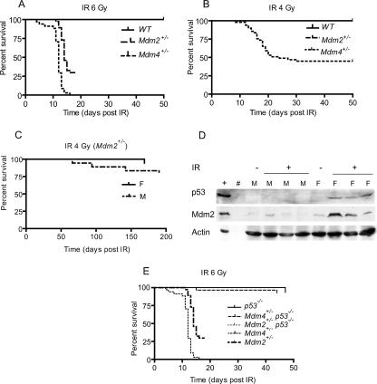
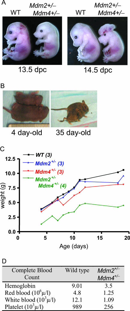
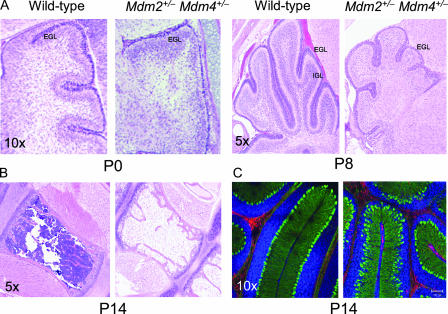
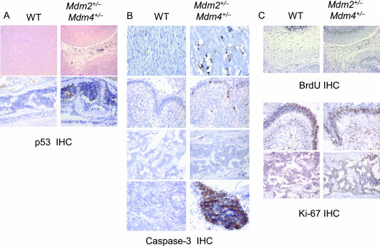
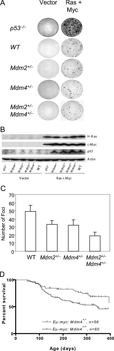
The tumor suppressor p53 is inactivated by multiple mechanisms that include mutations of the p53 gene itself and increased levels of the p53 inhibitors MDM2 and MDM4. Mice lacking Mdm2 or Mdm4 exhibit embryo-lethal phenotypes that are completely rescued by concomitant deletion of p53. Here we show that Mdm2 and Mdm4 haploinsufficiency leads to increased p53 activity, exhibited as increased sensitivity to DNA damage and decreased transformation potential. Moreover, in in vivo tumor development, Emu-myc Mdm4+/- mice show a delayed onset of B-cell lymphomas compared to Emu-myc mice. Additionally, Mdm2+/- Mdm4+/- double-heterozygous mice are not viable and exhibit defects in hematopoiesis and cerebellar development. The defects in Mdm2+/- Mdm4+/- mice are corrected by deletion of a single p53 allele. These findings highlight the exquisite sensitivity of p53 to Mdm2 and Mdm4 levels and suggest that some cell types may be more sensitive to therapeutic drugs that inhibit the Mdm-p53 interaction.


Figures

FIG. 1.
FIG. 1.
Radiosensitivity in Mdm2+/− and Mdm4+/− 5-week-old mice after a sublethal dose of whole-body IR. (A) Kaplan-Meier survival curves of age-matched wild-type (WT) (n = 65), Mdm2+/− (n = 37), and Mdm4+/− (n = 34) mice after 6 Gy of IR. (B) Kaplan-Meier survival curves of age-matched wild-type (n = 50), Mdm4+/− (n = 31), and Mdm2+/− (n = 39) mice after 4 Gy of IR. (C) Kaplan-Meier survival curves for Mdm2+/− mice by sex (18 females and 21 males). (D) Protein lysates from the thymuses of irradiated (+) and nonirradiated (−) male (M) and female (F) mice blotted with p53 and Mdm2 antibodies. Actin was used as a loading control. (E) Kaplan-Meier survival curves of Mdm2+/− (n = 37), Mdm4+/− (n = 34), Mdm2+/− p53−/− (n = 27), Mdm4+/− p53−/− (n = 28), and p53−/− (n = 9) mice.
FIG. 2.
FIG. 2.
Embryonic and postnatal phenotypes of Mdm2+/− Mdm4+/− mice. (A) Twenty percent of Mdm2+/− Mdm4+/− embryos show exencephaly associated with cleft palate or other neural tube defects. WT, wild type. (B) Delay in postnatal development of Mdm2+/− Mdm4+/− mice compared to wild-type mice. (C) Weights of mice with various genotypes during early postnatal development. (D) Complete blood counts for 13-day-old wild-type and Mdm2+/− Mdm4+/− pups.
FIG. 3.
FIG. 3.
Hypoplasia of bone marrow and cerebellum in Mdm2+/− Mdm4+/− mice. (A) Cerebellums from newborn (P0) and 8-day-old (P8) mice. Note the obvious hypoplasia of the EGL and the rudimentary internal granular layer (IGL) in P8 mice. (B) Bone marrow from the sternums of P14 Mdm2+/− Mdm4+/− mice. (C) Immunofluorescence staining showing the disorganization of Purkinje cells stained green for calbindin. Red, GFAP; blue, Topro3 nuclear stain.
FIG. 4.
FIG. 4.
p53 IHC, apoptosis, and proliferation assays. Paraffin-embedded sections from wild-type (WT) and Mdm2+/− Mdm4+/− mice were examined. (A) p53 IHC. (Upper row) Brains of 13.5-dpc embryos; (lower row) cranial bone marrow of 3-day-old pups. (B) For apoptosis, caspase-3 IHC of 13.5-dpc embryo brains (top row) and 1-day-old pup cerebellums (second row), cranial bone marrow (third row), and spleens (bottom row) is shown. (C) For proliferation, BrdU labeling of 13.5-dpc embryo brains (top row) and Ki-67 IHC of 3-day-old cerebellums (center row) and cranial bone marrow (bottom row) are shown. Note that the embryonic tissues do not compare due to developmental differences.
FIG. 5.
FIG. 5.
(A) Focus-forming assays with wild-type (WT), p53−/−, Mdm2+/−, Mdm4+/−, and Mdm2+/− Mdm4+/− MEFs. Early-passage MEFs were infected with two retrovirus vectors: one containing activated Ha-rasV12 and the second containing c-myc. p53−/− MEFs were used as a positive control. MEFs of different genotypes were also infected with vector alone as a negative control. (B) Western blotting of infected MEFs with c-myc and Ras antibodies. (C) Quantitative representation of the numbers of foci shown in panel A. (D) Delay of Myc-induced lymphomagenesis by Mdm4 haploinsufficiency. Kaplan-Meier survival curves of Eμ-myc Mdm4+/+ (n = 60) and Eμ-myc Mdm4+/− (n = 56) transgenic mice are shown.

References

    1. Adams, J. M., A. W. Harris, C. A. Pinkert, L. M. Corcoran, W. S. Alexander, S. Cory, R. D. Palmiter, and R. L. Brinster. 1985. The c-myc oncogene driven by immunoglobulin enhancers induces lymphoid malignancy in transgenic mice. Nature 318:533-538. - PubMed
    1. Alt, J. R., T. C. Greiner, J. L. Cleveland, and C. M. Eischen. 2003. Mdm2 haplo-insufficiency profoundly inhibits Myc-induced lymphomagenesis. EMBO J. 22:1442-1450. - PMC - PubMed
    1. Bond, G. L., W. Hu, E. E. Bond, H. Robins, S. G. Lutzker, N. C. Arva, J. Bargonetti, F. Barte, H. Taubert, P. Wuerl, K. Onel, L. Yip, S. J. Hwang, L. C. Strong, G. Lozano, and A. J. Levine. 2004. A single nucleotide polymorphism in the MDM2 promoter attenuates the p53 tumor suppressor pathway and accelerates tumor formation in humans. Cell 119:591-602. - PubMed
    1. Bond, G. L., W. Hu, and A. Levine. 2005. A single nucleotide polymorphism in the MDM2 gene: from a molecular and cellular explanation to clinical effect. Cancer Res. 65:5481-5484. - PubMed
    1. Danovi, D., E. Meulmeester, D. Pasini, D. Migliorini, M. Capra, R. Frenk, P. de Graaf, S. Francoz, P. Gasparini, A. Gobbi, K. Helin, P. G. Pelicci, A. G. Jochemsen, and J.-C. Marine. 2004. Amplification of Mdmx (or Mdm4) directly contributes to tumor formation by inhibiting p53 tumor suppressor activity. Mol. Cell. Biol. 24:5835-5843. - PMC - PubMed

Publication types