. 2007 Aug;75(8):4012-9.
doi: 10.1128/IAI.00645-07. Epub 2007 May 25.

Induction of nitric oxide synthase and activation of signaling proteins in Anopheles mosquitoes by the malaria pigment, hemozoin

Affiliations

Induction of nitric oxide synthase and activation of signaling proteins in Anopheles mosquitoes by the malaria pigment, hemozoin

Leyla Akman-Anderson et al. Infect Immun. 2007 Aug.

Abstract

Anopheles stephensi, a major vector for malaria parasite transmission, responds to Plasmodium infection by synthesis of inflammatory levels of nitric oxide (NO), which can limit parasite development in the midgut. We have previously shown that Plasmodium falciparum glycosylphosphatidylinositols (PfGPIs) can induce A. stephensi NO synthase (AsNOS) expression in the midgut epithelium in vivo in a manner similar to the manner in which cytokines and NO are induced by PfGPIs in mammalian cells. In mosquito cells, signaling by PfGPIs and P. falciparum merozoites is mediated through Akt/protein kinase B (Akt/PKB), the mitogen-activated protein kinase kinase DSOR1, and extracellular signal-regulated kinase (ERK). In mammalian cells, a second parasite factor, malaria pigment or hemozoin (Hz), signals NOS induction through ERK- and nuclear factor kappa B-dependent pathways and has been demonstrated to be a novel proinflammatory ligand for Toll-like receptor 9. In this study, we demonstrate that Hz can also induce AsNOS gene expression in immortalized A. stephensi and Anopheles gambiae cell lines in vitro and in A. stephensi midgut tissue in vivo. In mosquito cells, Hz signaling is mediated through transforming growth factor beta-associated kinase 1, Akt/PKB, ERK, and atypical protein kinase C zeta/lambda. Our results show that Hz is a prominent parasite-derived signal for Anopheles and that signaling pathways activated by PfGPIs and Hz have both unique and shared components. Together with our previous findings, our data indicate that parasite signaling of innate immunity is conserved in mosquito and mammalian cells.


Figures
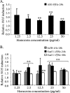
FIG. 1.
FIG. 1.
NOS expression is induced by sHz and PfHz in Anopheles cells. (A) ASE cells were stimulated with PfHz at concentrations of 1.25 to 50 μg/ml or with PBS as a control for 24 h (n = 3). (B) 4a3B cells were stimulated with sHz at concentrations of 1.25 to 50 μg/ml or with PBS as a control for 24 h (n = 4). Sua5.1 cells were treated with PfHz at the same concentrations for 24 and 48 h (n = 4). NOS expression values were divided by the values for PBS-treated controls to obtain relative induction levels. The error bars indicate standard errors of the means. Data within each treatment were analyzed using the Student t test. P values are represented as follows: two asterisks, P ≤ 0.01; one asterisk, P ≤ 0.05.
FIG. 2.
FIG. 2.
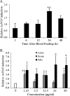
AsNOS expression is induced by PfHz in the midgut epithelium in vivo, and maximum induction occurs at 24 h pBM. (A) Midguts were dissected from A. stephensi at 1 to 48 h after blood feeding for analysis of AsNOS expression (n = 3). Artificial blood meals contained washed human RBCs and plasma supplemented with 50 μg/ml PfHz or PBS as a control. (B) Midguts were dissected from A. stephensi at 24 h after blood feeding with PfHz or hemin chloride at concentrations of 1.25 to 50 μg/ml or with an equivalent volume of PBS as a control (n = 3). Data were analyzed as described in the legend to Fig. 1 using the Student t test and are mean levels of relative AsNOS induction (NOS expression in PfHz- or hemin-fed groups divided by NOS expression in PBS-fed controls); the error bars indicate standard errors of the means. P values are represented as follows: two asterisks, P ≤ 0.01; one asterisk, P ≤ 0.05.
FIG. 3.
FIG. 3.
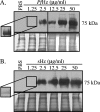
Both PfHz and sHz can activate TAK1. 4a3B cells were treated with PfHz (A) or sHz (B) at concentrations of 1.25 to 50 μg/ml or with equivalent volumes of PBS as a control for 10 min. After stimulation, cells were lysed and analyzed by Western blotting using anti-phospho-TAK1 antisera. An additional blot was used in panel A to show TAK1 phosphorylation in samples treated with 1.25 and 2.5 μg/ml PfHz. The insets show cross-reacting bands after prolonged film exposure. Equal loading of proteins was confirmed by Coomassie blue staining (bottom panels). The blots are representative of replicated Western blots from independent experiments.
FIG. 4.
FIG. 4.
Both PfHz and sHz can activate Akt/PKB. 4a3B cells were treated with PfHz (A) or sHz (B) at concentrations of 1.25 to 50 μg/ml or with equivalent volumes of PBS as a control for 10 min. After stimulation, cells were lysed and analyzed by Western blotting using anti-phospho-Akt/PKB antisera. The insets show cross-reacting bands after prolonged film exposure. Equal loading of proteins was confirmed by Coomassie blue staining (bottom panels). The blots are representative of replicated Western blots from independent experiments.
FIG. 5.
FIG. 5.
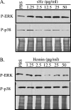
Hz and hemin are distinct signals. 4a3B cells were treated with sHz (A) or hemin chloride (B) at concentrations of 1.25 to 50 μg/ml or with equivalent volumes of PBS as a control for 10 min. After stimulation, cells were lysed and analyzed by Western blotting using anti-phospho-ERK (top panels) or anti-phospho-p38 antisera (middle panels). Equal loading of proteins was confirmed by Coomassie blue staining (bottom panels). The blots are representative of replicated Western blots from independent experiments.
FIG. 6.
FIG. 6.
sHz increases phosphorylation of cytosolic aPKCζ/λ. Sua5.1 cells were treated with 50 μg/ml sHz or an equivalent volume of PBS as a control for 5 min. After stimulation, cells were lysed and proteins were separated into cytosolic, membrane, and nuclear fractions. Equal amounts of proteins from the cellular compartments were analyzed by Western blotting using anti-phospho-aPKCζ/λ antisera. Equal loading of proteins was confirmed by Coomassie blue staining (bottom panel).

References

    1. An, H., Y. Yu, M. Zhang, H. Xu, R. Qi, X. Yan, S. Liu, W. Wang, Z. Guo, J. Guo, Z. Qin, and X. Cao. 2002. Involvement of ERK, p38 and NF-kappaB signal transduction in regulation of TLR2, TLR4 and TLR9 gene expression induced by lipopolysaccharide in mouse dendritic cells. Immunology 106:38-45. - PMC - PubMed
    1. Anstey, N. M., J. B. Weinberg, M. Y. Hassanali, E. D. Mwaikambo, D. Manyenga, M. A. Misukonis, D. R. Arnelle, D. Hollis, M. I. McDonald, and D. L. Granger. 1996. Nitric oxide in Tanzanian children with malaria: inverse relationship between malaria severity and nitric oxide production/nitric oxide synthase type 2 expression. J. Exp. Med. 184:557-567. - PMC - PubMed
    1. Arese, P., and E. Schwarzer. 1997. Malarial pigment (haemozoin): a very active ‘inert’ substance. Ann. Trop. Med. Parasitol. 91:501-516. - PubMed
    1. Avila, A., N. Silverman, M. T. Diaz-Meco, and J. Moscat. 2002. The Drosophila atypical protein kinase C-ref(2)p complex constitutes a conserved module for signaling in the toll pathway. Mol. Cell. Biol. 22:8787-8795. - PMC - PubMed
    1. Berner, R., W. Rudin, and H. Hecker. 1983. Peritrophic membranes and protease activity in the midgut of the malaria mosquito, Anopheles stephensi (Liston) (Insecta: Diptera) under normal and experimental conditions. J. Ultrastruct. Res. 83:195-204. - PubMed

Publication types

MeSH terms

LinkOut - more resources