. 2007 Aug;73(16):5338-46.
doi: 10.1128/AEM.02947-06. Epub 2007 May 25.

New insights into the colonization and release processes of Xenorhabdus nematophila and the morphology and ultrastructure of the bacterial receptacle of its nematode host, Steinernema carpocapsae

Affiliations

New insights into the colonization and release processes of Xenorhabdus nematophila and the morphology and ultrastructure of the bacterial receptacle of its nematode host, Steinernema carpocapsae

Holly Snyder et al. Appl Environ Microbiol. 2007 Aug.

Abstract

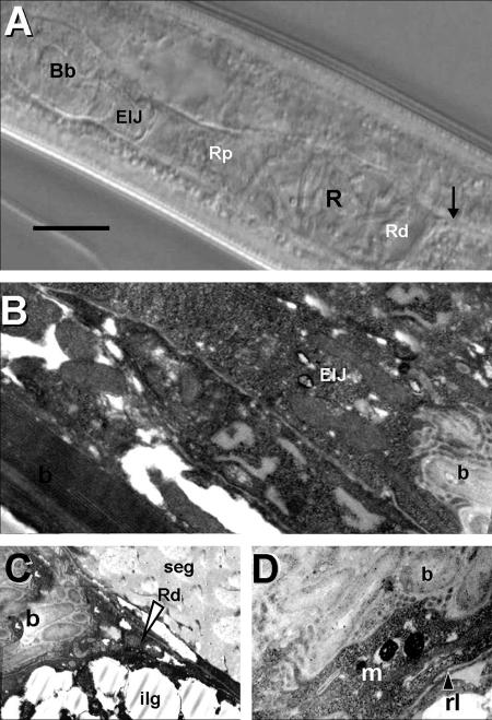
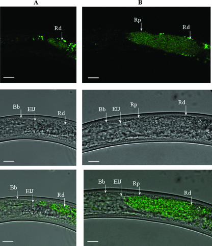
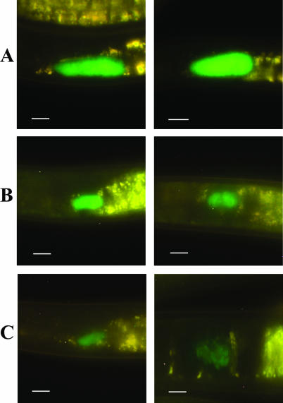
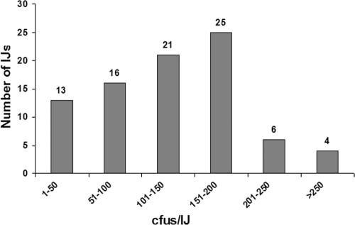
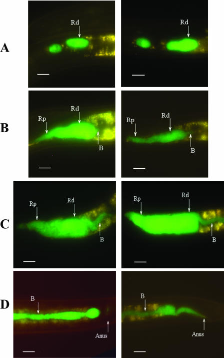
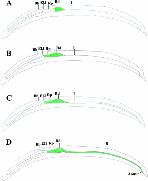
We present results from epifluorescence, differential interference contrast, and transmission electron microscopy showing that Xenorhabdus nematophila colonizes a receptacle in the anterior intestine of the infective juvenile (IJ) stage of Steinernema carpocapsae. This region is connected to the esophagus at the esophagointestinal junction. The process by which X. nematophila leaves this bacterial receptacle had not been analyzed previously. In this study we monitored the movement of green fluorescent protein-labeled bacteria during the release process. Our observations revealed that Xenorhabdus colonizes the distal region of the receptacle and that exposure to insect hemolymph stimulated forward movement of the bacteria to the esophagointestinal junction. Continued exposure to hemolymph caused a narrow passage in the distal receptacle to widen, allowing movement of Xenorhabdus down the intestine and out the anus. Efficient release of both the wild type and a nonmotile strain was evident in most of the IJs incubated in hemolymph, whereas only a few IJs incubated in nutrient-rich broth released bacterial cells. Incubation of IJs in hemolymph treated with agents that induce nematode paralysis dramatically inhibited the release process. These results suggest that bacterial motility is not required for movement out of the distal region of the receptacle and that hemolymph-induced esophageal pumping provides a force for the release of X. nematophila out of the receptacle and into the intestinal lumen.


Figures

FIG. 1.
FIG. 1.
DIC and TEM images of the bacterial receptacle of S. carpocapsae. (A) DIC image of colonized IJ receptacle (R) showing proximal (Rp) and distal (Rd) ends. The arrow indicates the connection between the distal receptacle and the intestine. The anterior end of the nematode is oriented to the left side of the panel. Bb, basal bulb. (B) TEM image of anterior portion of bacterial receptacle in connection with the EIJ. b, bacterial cells. (C) Posterior portion of bacterial receptacle showing end (arrowhead), Rd, and bacterial cells (b). ilg, intestinal lipid globule; seg, secretory-excretory gland. (D) Close-up of bacterial receptacle showing bacterial cells (b), receptacle lining (rl), and amorphous matrix (m). Bar, 15 μm.
FIG. 2.
FIG. 2.
Confocal image of X. nematophila cells (green) over DIC image. (A) Unstimulated IJ containing X. nematophila cells within the Rd. The anterior end of the nematode is oriented to the left side of the panels. Spots anterior and posterior to the receptacle are due to autofluorescent tissue of the nematode. (B) An IJ incubated in hemolymph for 24 h showing forward movement of X. nematophila from the Rd to the proximal receptacle (Rp). Top row, confocal images; middle row, DIC images; bottom row, overlay images. Bars, 10 μm.
FIG. 3.
FIG. 3.
Epifluorescent images of the level of colonization (high [A], medium [B], and low [C]) of the distal area of the receptacle in S. carpocapsae IJs. In all panels, the anterior end of the nematode is oriented to the left side of the panel. Left and right panels are images representative of the different levels of colonization. Bars, 10 μm.
FIG. 4.
FIG. 4.
Analysis of colonization levels by single nematode grindings. One hundred individual nematodes were individually homogenized to release bacteria from the receptacle.
FIG. 5.
FIG. 5.
Epifluorescent images of movement of X. nematophila in the receptacle of the IJ exposed to hemolymph. (A) Clusters of bacteria in the distal and proximal receptacle region. (B) Forward movement of cells to the EIJ region and initial movement of bacteria out of the posterior region of the distal receptacle. (C) Bacterial cells moving posteriorly out of the distal receptacle. (D) Bacterial cells moving down the intestine and out the anus. In all panels the anterior end of the nematode is oriented to the left. Left and right panels are images representative of the movement of bacteria in hemolymph-stimulated IJs. B, bacteria. Bars, 10 μm.
FIG. 6.
FIG. 6.
DIC images showing bacterial release process. Shown is the opening of the bacterial receptacle into the intestine. Notice the expansion of the intestinal lumen (il) and the migration of the bacterial cells (b) into this lumen. The anterior end of the nematode is oriented to the top of the panel. Bar, 6 μm.
FIG. 7.
FIG. 7.
Schematic representation of bacteria moving both forward towards the basal bulb and rearward out of the distal receptacle through the narrow passage and into the intestine. (A) The Rd colonized by X. nematophila in the unstimulated IJ. (B) IJs exposed to hemolymph begin pharyngeal pumping, and forward movement of bacteria commences. (C) Widening of the narrow passage in the distal receptacle allowing bacteria to move into the intestine. (D) Movement of bacteria down the intestine and out the anus. Bb, basal bulb; I, intestine; B, bacteria.

Similar articles

Cited by

References

    1. Akhurst, R. J. 1982. Antibiotic activity of Xenorhabdus spp., bacteria symbiotically associated with insect pathogenic nematodes of the families Heterorhabditidae and Steinernematidae. J. Gen. Microbiol. 128:3061-3065. - PubMed
    1. Akhurst, R. J. 1983. Neoaplectana species: specificity of association with bacteria of the genus Xenorhabdus. Exp. Parasitol. 55:258-263. - PubMed
    1. Akhurst, R. J., and N. E. Boemare. 1990. Biology and taxonomy of Xenorhabdus, p. 75-90. In R. Gaugler and H. K. Kaya (ed.), Entomopathogenic nematodes in biological control. CRC Press, Boca Raton, FL.
    1. Bird, A. F., and R. J. Akhurst. 1983. The nature of the intestinal vesicle in nematodes of the family Steinernematidae. Int. J. Parasitol. 16:511-518.
    1. Ciche, A. T., and J. C. Ensign. 2003. For the insect pathogen Photorhabdus luminescens, which end of the nematode is out? Appl. Environ. Microbiol. 69:1890-1897. - PMC - PubMed

Publication types

LinkOut - more resources