

In situ activity and spatial organization of anaerobic ammonium-oxidizing (anammox) bacteria in biofilms
- PMID: 17526785
- PMCID: PMC1951037
- DOI: 10.1128/AEM.00156-07
In situ activity and spatial organization of anaerobic ammonium-oxidizing (anammox) bacteria in biofilms
Abstract
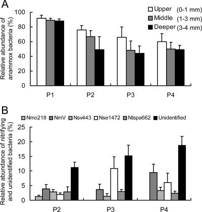
We investigated autotrophic anaerobic ammonium-oxidizing (anammox) biofilms for their spatial organization, community composition, and in situ activities by using molecular biological techniques combined with microelectrodes. Results of phylogenetic analysis and fluorescence in situ hybridization (FISH) revealed that "Brocadia"-like anammox bacteria that hybridized with the Amx820 probe dominated, with 60 to 92% of total bacteria in the upper part (<1,000 microm) of the biofilm, where high anammox activity was mainly detected with microelectrodes. The relative abundance of anammox bacteria decreased along the flow direction of the reactor. FISH results also indicated that Nitrosomonas-, Nitrosospira-, and Nitrosococcus-like aerobic ammonia-oxidizing bacteria (AOB) and Nitrospira-like nitrite-oxidizing bacteria (NOB) coexisted with anammox bacteria and accounted for 13 to 21% of total bacteria in the biofilms. Microelectrode measurements at three points along the anammox reactor revealed that the NH(4)(+) and NO(2)(-) consumption rates decreased from 0.68 and 0.64 micromol cm(-2) h(-1) at P2 (the second port, 170 mm from the inlet port) to 0.30 and 0.35 micromol cm(-2) h(-1) at P3 (the third port, 205 mm from the inlet port), respectively. No anammox activity was detected at P4 (the fourth port, 240 mm from the inlet port), even though sufficient amounts of NH(4)(+) and NO(2)(-) and a high abundance of anammox bacteria were still present. This result could be explained by the inhibitory effect of organic compounds derived from biomass decay and/or produced by anammox and coexisting bacteria in the upper parts of the biofilm and in the upstream part of the reactor. The anammox activities in the biofilm determined by microelectrodes reflected the overall reactor performance. The several groups of aerobic AOB lineages, Nitrospira-like NOB, and Betaproteobacteria coexisting in the anammox biofilm might consume a trace amount of O(2) or organic compounds, which consequently established suitable microenvironments for anammox bacteria.
Figures


Similar articles
-
Development of high-rate anaerobic ammonium-oxidizing (anammox) biofilm reactors.Water Res. 2007 Apr;41(8):1623-34. doi: 10.1016/j.watres.2007.01.050. Epub 2007 Mar 12. Water Res. 2007. PMID: 17350073
-
Microbial composition and structure of a rotating biological contactor biofilm treating ammonium-rich wastewater without organic carbon.Microb Ecol. 2003 May;45(4):419-32. doi: 10.1007/s00248-002-2037-5. Epub 2003 Apr 22. Microb Ecol. 2003. PMID: 12704553
-
Induced cooperation between marine nitrifiers and anaerobic ammonium-oxidizing bacteria by incremental exposure to oxygen.Syst Appl Microbiol. 2010 Nov;33(7):407-15. doi: 10.1016/j.syapm.2010.08.003. Epub 2010 Oct 16. Syst Appl Microbiol. 2010. PMID: 20956064
-
Global impact and application of the anaerobic ammonium-oxidizing (anammox) bacteria.Biochem Soc Trans. 2006 Feb;34(Pt 1):174-8. doi: 10.1042/BST0340174. Biochem Soc Trans. 2006. PMID: 16417514 Review.
-
Biomarkers for in situ detection of anaerobic ammonium-oxidizing (anammox) bacteria.Appl Environ Microbiol. 2005 Apr;71(4):1677-84. doi: 10.1128/AEM.71.4.1677-1684.2005. Appl Environ Microbiol. 2005. PMID: 15811989 Free PMC article. Review. No abstract available.
Cited by
-
Abundance and diversity of nitrogen-removing microorganisms in the UASB-anammox reactor.PLoS One. 2019 Apr 22;14(4):e0215615. doi: 10.1371/journal.pone.0215615. eCollection 2019. PLoS One. 2019. PMID: 31009503 Free PMC article.
-
Metabolic network analysis reveals microbial community interactions in anammox granules.Nat Commun. 2017 May 31;8:15416. doi: 10.1038/ncomms15416. Nat Commun. 2017. PMID: 28561030 Free PMC article.
-
Absolute quantification of individual biomass concentrations in a methanogenic coculture.AMB Express. 2014 Apr 12;4:35. doi: 10.1186/s13568-014-0035-x. eCollection 2014. AMB Express. 2014. PMID: 24949269 Free PMC article.
-
Community structure and in situ activity of nitrifying bacteria in Phragmites root-associated biofilms.Microbes Environ. 2012;27(3):242-9. doi: 10.1264/jsme2.me11314. Epub 2012 Mar 23. Microbes Environ. 2012. PMID: 22446303 Free PMC article.
-
Dynamics of microbial community structure of and enhanced biological phosphorus removal by aerobic granules cultivated on propionate or acetate.Appl Environ Microbiol. 2011 Nov;77(22):8041-51. doi: 10.1128/AEM.05738-11. Epub 2011 Sep 16. Appl Environ Microbiol. 2011. PMID: 21926195 Free PMC article.
References
-
- Altschul, S. F., W. Gish, W. Miller, E. W. Myers, and D. J. Lipman. 1990. Basic local alignment search tool. J. Mol. Biol. 215:403-410. - PubMed
-
- Andrussow, L. 1969. Diffusion, p. 513-727. In H. Borchers, H. Hauser, K. H. Hellwege, K. Schafer, and E. Schmidt (ed.), Landolt-Bornstein zahlenwerte und functionen, vol. II/5a. Springer, Berlin, Germany.
-
- Daims, H., A. Brühl, R. Amann, K.-H. Schleifer, and M. Wagner. 1999. The domain-specific probe EUB338 is insufficient for the detection of all bacteria: development and evaluation of a more comprehensive probe set. Syst. Appl. Microbiol. 22:434-444. - PubMed
Publication types
MeSH terms
Substances
Associated data
- Actions
- Actions
- Actions
- Actions
- Actions
- Actions
LinkOut - more resources
Full Text Sources
Molecular Biology Databases
