. 2007 Sep 15;110(6):2132-9.
doi: 10.1182/blood-2007-04-083873. Epub 2007 May 25.

Cytoplasmic remodeling of erythrocyte raft lipids during infection by the human malaria parasite Plasmodium falciparum

Affiliations

Cytoplasmic remodeling of erythrocyte raft lipids during infection by the human malaria parasite Plasmodium falciparum

Sean C Murphy et al. Blood. .

Abstract

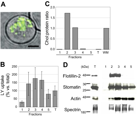
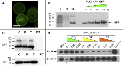
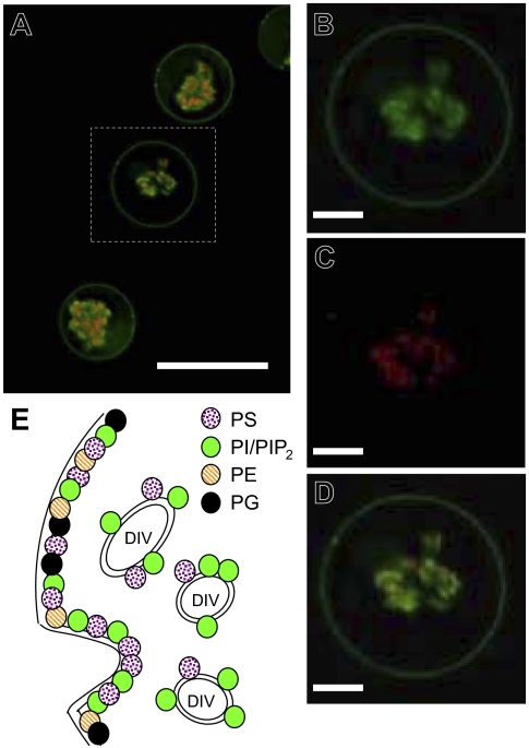
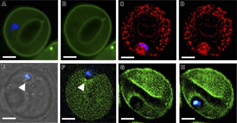
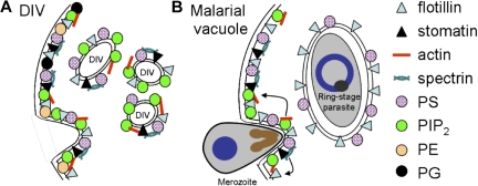
Studies of detergent-resistant membrane (DRM) rafts in mature erythrocytes have facilitated identification of proteins that regulate formation of endovacuolar structures such as the parasitophorous vacuolar membrane (PVM) induced by the malaria parasite Plasmodium falciparum. However, analyses of raft lipids have remained elusive because detergents interfere with lipid detection. Here, we use primaquine to perturb the erythrocyte membrane and induce detergent-free buoyant vesicles, which are enriched in cholesterol and major raft proteins flotillin and stomatin and contain low levels of cytoskeleton, all characteristics of raft microdomains. Lipid mass spectrometry revealed that phosphatidylethanolamine and phosphatidylglycerol are depleted in endovesicles while phosphoinositides are highly enriched, suggesting raft-based endovesiculation can be achieved by simple (non-receptor-mediated) mechanical perturbation of the erythrocyte plasma membrane and results in sorting of inner leaflet phospholipids. Live-cell imaging of lipid-specific protein probes showed that phosphatidylinositol (4,5) bisphosphate (PIP(2)) is highly concentrated in primaquine-induced vesicles, confirming that it is an erythrocyte raft lipid. However, the malarial PVM lacks PIP(2), although another raft lipid, phosphatidylserine, is readily detected. Thus, different remodeling/sorting of cytoplasmic raft phospholipids may occur in distinct endovacuoles. Importantly, erythrocyte raft lipids recruited to the invasion junction by mechanical stimulation may be remodeled by the malaria parasite to establish blood-stage infection.


Figures

Figure 1
Figure 1
Characteristics of primaquine-induced endovesicles. (A) Micrograph of a live mature erythrocyte containing LY+ primaquine-induced endovesicles. Single merged optical section of differential interference contrast (DIC) and green fluorescent (LY) channels; 60×/1.42 oil objective (Olympus). Scale bar equals 3 μm. (B) Association of LY with membrane fractions 1 through 5 from primaquine-treated erythrocytes separated by sucrose density centrifugation and quantified by immunoblotting with anti-LY antibodies and densitometry. Fraction numbers are indicated (1, top; 5, bottom). Bars show total LY signal in each fraction divided by total LY signal in the starting material (T) expressed as a percentage. Error bars indicates standard deviation of duplicate measurements. (C) Cholesterol-protein ratios of endovesicle fractions 1 through 5. Cholesterol and protein levels were measured and ratios calculated as described for erythrocyte DRMs; hemoglobin-free erythrocyte membranes were used as reference values. T indicates starting material; WM, hemoglobin-free white membranes. (D) Immunblotting of erythrocyte endovesicle membrane fractions using specific antisera for the indicated human proteins (flotillin-2, stomatin, actin, spectrin) and specific secondary antibodies. T indicates total lysate. Fractions 2 and 3 were enriched in the raft protein flotillin-2, mildly enriched for human stomatin, and contained lesser amounts of cytoskeletal proteins actin and spectrin.
Figure 2
Figure 2
The GFP-PLCδ1 PH domain preferentially binds PIP2 on the cytoplasmic face of the plasma membrane of loaded-erythrocyte ghosts. (A) Single optical sections of fluorescence micrographs (60×/1.42 oil objective [Olympus]) of erythrocyte ghosts loaded with recombinant GFP-PLCδ1 PH domain (5 μM) protein. Scale bar equals approximately 5 μm. (B) Membrane association of GFP-PLCδ1 PH domain in loaded ghosts. Ghosts loaded with GFP-PLCδ1 PH domain were lysed and fractionated into membrane and cytoplasmic fractions by centrifugation, separated by SDS-PAGE, and analyzed for recombinant protein by anti-GFP immunoblotting. T, total PLCδ1 PH domain–loaded ghost lysate; C, cytoplasmic fraction; M, membrane fraction; 5 × 10⁷ cells per lane; protein loading equal by Ponceau staining. Purified GFP-PLCδ1 standard is shown at right; nanogram scale. (C) Protease protection assay of GFP-PLCδ1 PH domain in loaded ghosts. Ghosts loaded with GFP-PLCδ1 PH domain were incubated in buffer with or without proteinase K and/or 1% Triton X-100 detergent, then separated and analyzed as in panel B. In addition to anti-GFP immunoblotting, control antibodies to erythrocyte flotillin-1 (flot1; completely protease protected) were also used. C, control; P, proteinase K; T, Triton X-100; P/T, proteinase K and Triton X-100; 2 × 10⁷ cells per lane; protein loading equal by Ponceau staining. (D) In vitro liposome association of GFP PLCδ1 PH domains. Recombinant GFP PLCδ1 PH domain fusion protein was incubated with purified liposomes containing PIP2, PI4P, or PA, separated into a liposome-containing pellet (P) and liposome-depleted supernatant (S) fractions by ultracentrifugation, and immunoblotted for GFP as described in the other panels. Numbers represent the relative ratios of DMPC to PIP2, PI4P, or PA in liposomes (0.1 = 1:20; 0.5 = 1:40; 0.025 = 1:80). *Lane with most PH domain binding of PIP2-containing liposomes.
Figure 3
Figure 3
The GFP-PLCδ1 PH domain accumulates on drug-induced endovesicles. (A-D) Fluorescence micrographs (60×/1.42 oil objective [Olympus]) of erythrocyte ghosts loaded with GFP- PLCδ1 PH domain (panel B; green) and treated with primaquine in the presence of 70-kDa rhodamine dextran (panel C; red). Rhodamine dextran accumulated inside endovesicles, and the GFP-PLCδ1 PH domain colocalized to the same region as rhodamine dextran. Merge shown in panels A (bar equals 10 μm) and D. Dashed box in panel A denotes cell magnified in panels B through D. Scale bar equals 3 μm (panels B-D). (E) Model of erythrocyte lipid uptake and exclusion in drug-induced vesicles (DIVs). Based on mass spectrometry and immunofluorescence results, DIVs appear to undergo complex but ordered lipid sorting during their primaquine-induced formation. In particular, whereas PE and PG are absent from isolated endovesicles, PS and especially PI phospholipids traffic to the endovesicle membranes. With respect to PIP2, this is quantitatively apparent by mass spectrometry and fluorescence microscopy. DIVs may consist of smaller, dynamic raft microdomains containing PIP2 and PS. The extent to which the ratio of cytoskeletal proteins such as spectrin and actin are changed in DIV as well as the presence of junctional complex proteins like 4.1 in DIV have not yet been established.
Figure 4
Figure 4
PIP2 is absent from malaria-induced vacuoles. (A,B) Fluorescence micrographs of a GFP-PLCδ1 PH domain–loaded ghost (green) infected by a young ring-stage malaria parasite. The parasite nucleus is stained with blue Hoechst 33342 as shown in the merged image. Scale bar equals 2 μm. (C,D). Erythrocyte ghost loaded with Alexa-Fluor 594–labeled annexin V (red) containing a Hoechst-stained intracellular ring-stage parasite as shown in the merged image. Scale bar equals 2 μm. (E,F). Merozoite (blue) attached to the surface (as shown by light microscopy) (E) and indenting (arrowhead) the membrane of an erythrocyte ghost loaded with GFP-PLCδ1 PH domain (F). Scale bar equals 2 μm. (G-H). An erythrocyte ghost loaded with GFP-PLCδ1 PH domain (G) containing a recently invaded young ring-stage parasite (H) (merge with Hoechst stain). Images acquired with a 100×/1.35 oil objective (Olympus).
Figure 5
Figure 5
Model of lipid and protein uptake into the erythrocyte DIV and the malarial vacuole. (A) The erythrocyte membrane contains a variety of phospholipids (PS, PI/PIP2, PE, PG, etc) and raft (stomatin, flotillins) and nonraft (actin, spectrin) proteins, most of which are taken up into DIV membranes. However, PE- and PG-type phospholipids are specifically excluded from these DIVs. Triangles represent membrane raft proteins flotillin-1 and flotillin-2 (light blue) and stomatin (black). Red bars and blue curved bars represent cytoskeletal actin and spectrin, respectively. Circles represent phospholipids PS (purple), PIP2 (green), PE (yellow), or PG (black). (B) Model of lipid and protein uptake into the malarial vacuolar membrane. Although the merozoite-induced indentation of the nascent vacuole may be remarkably similar to that of DIVs, the fully formed vacuole appears specifically enriched in flotillins, some additional raft proteins (not shown), and host PS. Unlike DIVs, the malarial vacuolar membrane is devoid of cytoskeleton, PIP2, and membrane raft stomatin; arrows depict the selective departure of actin, PIP2, stomatin, and spectrin from the forming malarial vacuole. This selectivity may reflect the combined contribution of erythrocyte-dependent molecular sorting on the basis of membrane curvature and lipid composition as well as potential contributions from parasite-encoded factors that serve to modify the vacuolar membrane during malarial invasion. PE and PG are not shown in panel B. In panel A, a flippase activity is implicated to keep PS cytoplasmically oriented in erythrocytes and DIVs. In malarial invasion (panel B), PS is not exposed at the infected erythrocyte membrane during PVM formation; it remains unknown whether PS is lumenal in the PVM. Our result that PS is detected on the cytoplasmic face of the vacuole is the first clear evidence of PS distribution in the PVM.

Similar articles

Cited by

References

    1. Dvorak JA, Miller LH, Whitehouse WC, Shiroishi T. Invasion of erythrocytes by malaria merozoites. Science. 1975;187:748–750. - PubMed
    1. Holder AA, Blackman MJ, Borre M, et al. Malaria parasites and erythrocyte invasion. Biochem Soc Trans. 1994;22:291–295. - PubMed
    1. Miller LH, Baruch DI, Marsh K, Doumbo OK. The pathogenic basis of malaria. Nature. 2002;415:673–679. - PubMed
    1. Murphy SC, Samuel BU, Harrison T, et al. Erythrocyte detergent-resistant membrane proteins: their characterization and selective uptake during malarial infection. Blood. 2004;103:1920–1928. - PubMed
    1. Samuel BU, Mohandas N, Harrison T, et al. The role of cholesterol and glycosylphosphatidylinositol-anchored proteins of erythrocyte rafts in regulating raft protein content and malarial infection. J Biol Chem. 2001;276:29319–29329. - PubMed

Publication types

MeSH terms