. 2007 May;3(5):e94.
doi: 10.1371/journal.pcbi.0030094. Epub 2007 Apr 12.

Prediction of functional sites based on the fuzzy oil drop model

Affiliations

Prediction of functional sites based on the fuzzy oil drop model

Michał Bryliński et al. PLoS Comput Biol. 2007 May.

Abstract

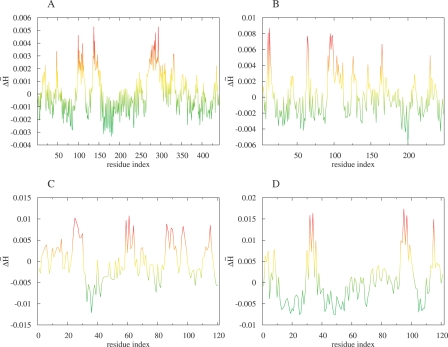
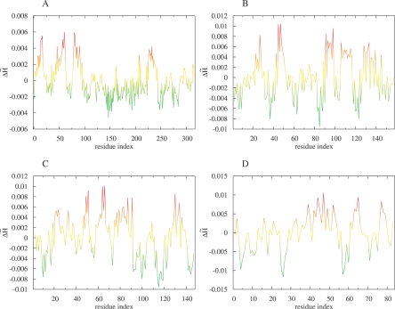
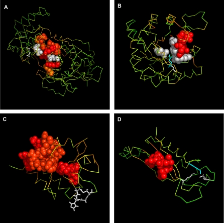
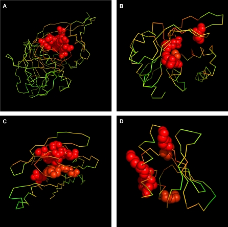
A description of many biological processes requires knowledge of the 3-D structure of proteins and, in particular, the defined active site responsible for biological function. Many proteins, the genes of which have been identified as the result of human genome sequencing, and which were synthesized experimentally, await identification of their biological activity. Currently used methods do not always yield satisfactory results, and new algorithms need to be developed to recognize the localization of active sites in proteins. This paper describes a computational model that can be used to identify potential areas that are able to interact with other molecules (ligands, substrates, inhibitors, etc.). The model for active site recognition is based on the analysis of hydrophobicity distribution in protein molecules. It is shown, based on the analyses of proteins with known biological activity and of proteins of unknown function, that the region of significantly irregular hydrophobicity distribution in proteins appears to be function related.


Conflict of interest statement

Competing interests. The authors have declared that no competing interests exist.

Figures

Figure 1
Figure 1. Profile Plots of Hydrophobicity Deviation Δ H~ per Amino Acid Obtained for Exemplary Proteins of Known Function
(A) Phosphomannose isomerase and (B) triosophosphate isomerase are examples of the high agreement with experimental data. (C) Protein disulfide isomerase and (D) 7,8-dihydroneopterin aldolase are examples of low agreement. The common color scale is introduced: red, high Δ H~; yellow, middle Δ H~; green, low and negative Δ H~.
Figure 2
Figure 2. Profile Plots of Hydrophobicity Deviation Δ H~ per Amino Acid Obtained for Exemplary Proteins of Unknown Function
The protein identified in the genome of Pseudomonas aeruginosa (A) and the protein identified in the genome of Thermotoga maritima (B) are examples representing close localization of residues of high Δ H~. The protein originated in the Thermus thermophilus genome (C) and the protein originated the Staphylococcus aureus genome (D) are examples of dispersed localization of residues representing high Δ H~. The common color scale (same as in Figure 1) is introduced: (low and negative Δ H~ proteins need additional analysis of their specificity).
Figure 3
Figure 3. The 3-D Representation of Proteins of Known Biological Activity with Binding Site Recognized
Phosphomannose isomerase (A) and triosophosphate isomerase (B) are examples of the high agreement with experimental data. Protein disulfide isomerase (C) and 7,8-dihydroneopterin aldolase (D) are examples of low agreement. Amino acids indicated by FOD as belonging to the binding cavity are in CPK form. The common color scale (same as in Figure 1) is introduced. The white color denotes the experimentally verified amino acids as active site (identification according to the CSA database).
Figure 4
Figure 4. The 3-D Representation of Proteins of Unknown Biological Activity with Binding Site Recognized
The protein identified in the Pseudomonas aeruginosa genome (A) and the protein identified in the Thermotoga maritima genome (B) are examples representing close localization of residues of high Δ H~. The protein originated in the Thermus thermophilus genome (C) and the protein originated in the Staphylococcus aureus genome (D) are examples of dispersed localization of residues representing high Δ H~. The common color scale (same as in Figure 1) is introduced.

References

    1. Burley SK, Almo SC, Bonanno JB, Capel M, Chance MR, et al. Structural genomics: Beyond the human genome project. Nat Genet. 1999;23:151–157. - PubMed
    1. Chance MR, Bresnick AR, Burley SK, Jiang JS, Lima CD, et al. Structural genomics: A pipeline for providing structures for the biologist. Protein Sci. 2002;11:723–738. - PMC - PubMed
    1. Goulding CW, Apostol M, Anderson DH, Gill HS, Smith CV, et al. The TB structural genomics consortium: Providing a structural foundation for drug discovery. Curr Drug Targets Infect Disord. 2002;2:121–141. - PubMed
    1. Chandonia JM, Earnest TN, Brenner SE. Structural genomics and structural biology: Compare and contrast. Genome Biol. 2004;5:343. - PMC - PubMed
    1. Zvelebil MJ, Sternberg MJ. Analysis and prediction of the location of catalytic residues in enzymes. Protein Eng. 1988;2:127–138. - PubMed

Publication types