. 2007 May 25;3(5):e66.
doi: 10.1371/journal.ppat.0030066.

Accumulation of pathological prion protein PrPSc in the skin of animals with experimental and natural scrapie

Affiliations

Accumulation of pathological prion protein PrPSc in the skin of animals with experimental and natural scrapie

Achim Thomzig et al. PLoS Pathog. .

Abstract

Prion infectivity and its molecular marker, the pathological prion protein PrP(Sc), accumulate in the central nervous system and often also in lymphoid tissue of animals or humans affected by transmissible spongiform encephalopathies. Recently, PrP(Sc) was found in tissues previously considered not to be invaded by prions (e.g., skeletal muscles). Here, we address the question of whether prions target the skin and show widespread PrP(Sc) deposition in this organ in hamsters perorally or parenterally challenged with scrapie. In hamsters fed with scrapie, PrP(Sc) was detected before the onset of symptoms, but the bulk of skin-associated PrP(Sc) accumulated in the clinical phase. PrP(Sc) was localized in nerve fibres within the skin but not in keratinocytes, and the deposition of PrP(Sc) in skin showed no dependence from the route of infection and lymphotropic dissemination. The data indicated a neurally mediated centrifugal spread of prions to the skin. Furthermore, in a follow-up study, we examined sheep naturally infected with scrapie and detected PrP(Sc) by Western blotting in skin samples from two out of five animals. Our findings point to the skin as a potential reservoir of prions, which should be further investigated in relation to disease transmission.


Conflict of interest statement

Competing interests. The authors have declared that no competing interests exist.

Figures

Figure 1
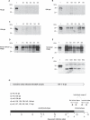
Figure 1. Time-Course of PrPSc Deposition in Skin Tissue
(A–E) Western blot detection of PrP27–30, the protease-resistant core of PrPSc, extracted from different skin samples of hamsters orally challenged with 263K scrapie and sacrificed at the following time-points after infection: (A) 70 dpi, (B) 100 dpi, (C) 130 dpi, (D) at the onset of clinical signs for scrapie (138–146 dpi), and (E) at the terminal stage of disease (157–171 dpi). Lanes with test samples: S1, skin sample from hindlimb; S2, skin sample from forelimb; S3, skin sample from back; S4, skin sample from abdomen; S5, skin sample from head. Lanes with control samples: 1, proteinase K–digested brain homogenate from terminally ill 263K scrapie hamsters containing 1 × 10⁻⁷ g brain tissue. Representative results are shown for each stage of incubation. Substantial individual variation was observed at 130 dpi, with two of five and three of five animals displaying findings as in (C) in the Western blot on the left-hand side or the Western blot on the right-hand side, respectively. (F) Lanes S1d–S5d: Same samples as in S1–S5 of (E) but deglycosylated with PNGaseF. (A–F) Amounts of tissue represented in lanes: (A) S1, 43 mg; S2, 52 mg; S3, 68 mg; S4, 58 mg; S5, 73 mg; (B) S1, 78 mg; S2, 44 mg; S3, 63 mg; S4, 67 mg; S5, 50 mg; ([C], Western blot on the left side) S1, 42 mg; S2, 76 mg; S3, 61 mg; S4, 58 mg; S5, 73 mg; ([C], Western blot on the right side) S1, 51 mg; S2, 63 mg; S3, 70 mg; S4, 87 mg; S5, 54 mg; (D) S1, 63 mg; S2, 68 mg; S3, 90 mg; S4, 50 mg; S5, 68 mg; (E) S1, 55 mg; S2, 73 mg; S3, 80 mg; S4, 88 mg; S5, 70 mg; (F) S1d, 12 mg; S2d, 14 mg; S3d, 19 mg; S4d, 12 mg; S5d, 20 mg. (G) Time-scale displaying the mean incubation period and the pre-clinical and clinical phases of incubation of hamsters orally infected with 263K scrapie. Small vertical arrows indicate time-points at which animals were tested for PrPSc in skin samples.
Figure 2
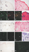
Figure 2. Location of PrPSc within the Skin of Hamsters Orally Infected with Scrapie
(A–H) Topographical localisation of PrPSc in sections of skin samples from the snout (A and B) and the forelimb (G and H); (A and G) PET blots, (B and H) H&E staining. PrPSc was detected in free nerve endings of the subepidermal plexus on the border of the epidermis to the dermis ([A], [B], [G], and [H], arrows), in fibres of the subepidermal, the deep cutaneous and the subcutaneous plexus, in circular and longitudinal fibres of the follicular neural network of the hair ([A], [B], and [G], arrowheads), in the hair follicle isthmus ([G]; rhombus), and in small intradermal striated fibres of mimic muscles ([A and B], asterisks). (C–F) Visualisation of PrPSc and nerve fibres in the neural network of hair follicles by fluorescence microscopy (skin sample from the abdomen). Co-localisation of PrPSc (C) with nerve fibres labelled by using an anti–S-100 protein antibody against Schwann cells (D). (E) Merged figure from micrographs (C and D). (F) Adjacent section to (C), stained with H&E. (I–K) Visualisation of PrPSc and nerve fibres in the cutaneous plexus by fluorescence microscopy (skin sample form the snout). Co-localisation of PrPSc (I) with nerve fibres labelled by using the anti-neurofilament antibody SMI 31 (J). (K) Merged figure from micrographs (I and J). (L) Adjacent section to (I), stained with H&E. The box indicates the region used for the immunofluorescence stainings in (I–K). (M and N) Control skin samples from the forelimb of a hamster perorally mock-challenged with normal hamster brain homogenate; PET blot (M) and fluorescence microscopy for PrP and neurofilament (N). Scale bars = 200 μm for (B, F, H, and M), 50 μm for (K and L), and 25μm for (N). Same scale bars as displayed in (B), (F), (H), and (K) apply to (A), (C–E), (G), and (I and J), respectively.
Figure 3
Figure 3. PrPSc Routing to the Skin and to Components of the Lymphoreticular System of Hamsters Challenged via Different Routes with 263K Scrapie Agent
(A) Western blot detection of PrP27–30, the protease-resistant core of PrPSc, in skin specimens from terminally ill scrapie hamsters. Lanes 1, 2, and 3: skin samples from orally mock-infected control hamsters, spiked before extraction with 1 × 10⁻⁶ g, 5 × 10⁻⁶ g, or 1 × 10⁻⁵ g of brain homogenate from terminally ill 263K hamsters. Lanes 4 and 5: skin samples from hindlimbs and forelimbs of hamsters orally infected with scrapie brain homogenate. Lanes 6 and 7: skin samples from hindlimbs and forelimbs of hamsters intracerebrally infected with scrapie brain homogenate. Lanes 8 and 9: skin samples from hindlimbs and forelimbs of hamsters infected by implantation of s.w. contaminated with scrapie agent. Lanes 10 and 11: skin samples from hindlimbs and forelimbs of hamsters infected peripherally by f.p. inoculation of scrapie brain homogenate. Lanes 12 and 13: skin samples from hindlimbs and forelimbs of hamsters orally mock-infected with normal brain homogenate. Amounts of tissue represented in lanes: 1, 53mg; 2, 58 mg; 3, 68 mg; 4, 68 mg; 5, 75 mg; 6, 78 mg; 7, 64 mg; 8, 69 mg; 9, 60 mg; 10, 62 mg; 11, 73 mg; 12, 61 mg; 13, 58 mg. (B) Western blot detection of PrP27–30 in spleens and selected lymph nodes from terminally ill scrapie hamsters. Lanes 1 and 5: proteinase K-digested brain homogenate from terminally ill scrapie hamsters, containing 1 × 10⁻⁷ g brain tissue. Lanes 2–4: spleen samples from p.o.- (2), s.w.-, (3) and i.c.-infected (4) hamsters. Lanes 6–8: mesenteric lymph node samples from p.o.- (6), s.w.-, (7) and i.c.-infected (8) hamsters. Amounts of tissue represented in lanes: 2, 40 mg; 3, 45 mg; 4, 41 mg; 6, 6 mg; 7, 8 mg; 8, 6 mg.
Figure 4
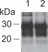
Figure 4. Western Blot Detection of PrP27–30 in Skin Specimens of Hamsters Intracerebrally Challenged with BSE-H Agent
Lanes 1 and 2: skin samples from hindlimb and forelimb of a BSE-infected hamster. Amounts of tissue represented in lanes: 1, 43 mg; 2, 58 mg.
Figure 5
Figure 5. PrPSc in Skin Samples from Different Body Areas of Sheep Naturally Infected with Scrapie
Western blot detection of PrP27–30, the protease-resistant core of PrPSc, using the anti-PrP antibodies ICSM-18 (lanes 1–8, 10–12) and P4 (lane 9). Samples are from sheep Sc3 (lanes 1–5), sheep Sc5 (lanes 6–9), and from an uninfected control sheep. Lanes 1 and 2: different amounts of brain homogenate (0.5 mg/lane and 0.1 mg/lane of midbrain tissue, respectively) from sheep Sc3. Lane 3: tonsil sample. Lane 4: tonsil sample after deglycosylation with PNGaseF. Lane 5: skin sample from the inguinal region. Lane 6: skin sample from the perianal region (scratching area). Lane 7: skin sample from the same region as in lane 6 after deglycosylation with PNGaseF. Lane 8: skin sample from the snout. Lane 9: skin sample from the same region as in lane 8, but detected with the antibody P4. Lanes 10–12: negative controls of skin samples from different body regions of an uninfected sheep. Amounts of tissue represented in lanes: 1, 0.5 mg; 2, 0.1 mg; 3, 7 mg; 4, 2 mg; 5, 88 mg; 6, 89 mg; 7, 23 mg; 8, 90 mg; 9, 92 mg; 10, 80 mg; 11, 87 mg; 12, 94 mg.

Similar articles

Cited by

References

    1. Prusiner SB. Prions. Proc Natl Acad Sci U S A. 1998;95:13363–13383. - PMC - PubMed
    1. Beekes M, McBride P. The spread of prions through the body in naturally acquired transmissible spongiform encephalopathies. FEBS J. 2007;274:588–605. - PubMed
    1. Beekes M, Baldauf E, Diringer H. Sequential appearance and accumulation of pathognomonic markers in the central nervous system of hamsters orally infected with scrapie. J Gen Virol. 1996;77:1925–1934. - PubMed
    1. van Keulen LJ, Vromans ME, van Zijderveld FG. Early and late pathogenesis of natural scrapie infection in sheep. APMIS. 2002;110:23–32. - PubMed
    1. Ironside JW. Pathology of variant Creutzfeldt-Jakob disease. Arch Virol Suppl. 2000. pp. 143–151. - PubMed

Publication types