. 2007 Sep 30;366(2):277-92.
doi: 10.1016/j.virol.2007.04.023. Epub 2007 May 24.

Ubiquitination and proteasomal degradation of interferon regulatory factor-3 induced by Npro from a cytopathic bovine viral diarrhea virus

Affiliations

Ubiquitination and proteasomal degradation of interferon regulatory factor-3 induced by Npro from a cytopathic bovine viral diarrhea virus

Zihong Chen et al. Virology. .

Abstract

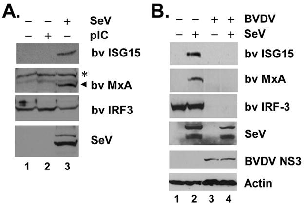
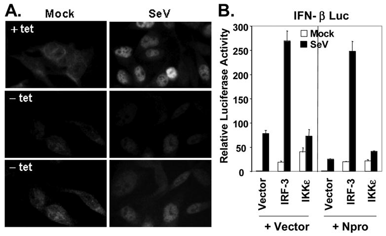
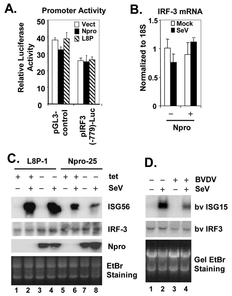
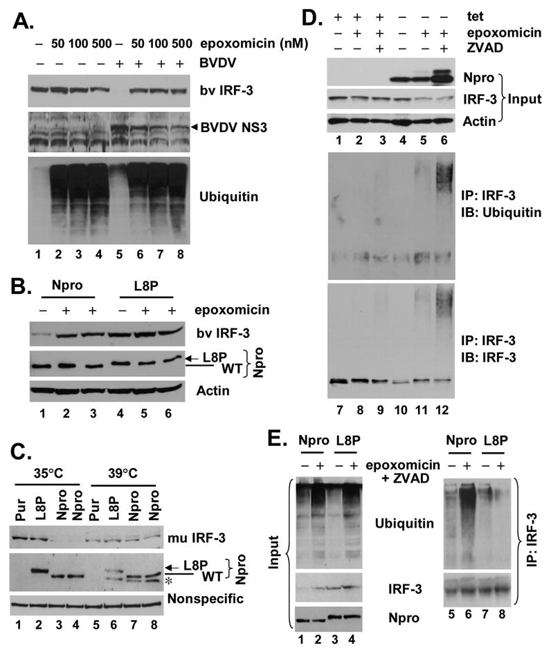
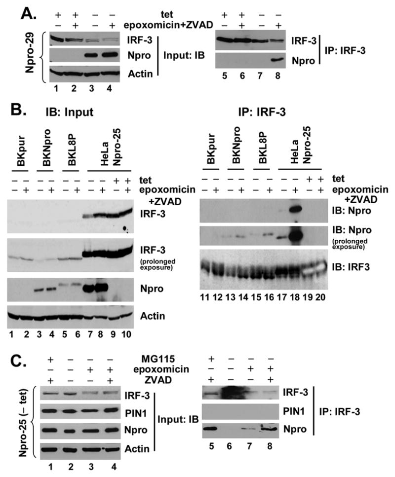
The pathogenesis of bovine viral diarrhea virus (BVDV) infections is complex and only partly understood. It remains controversial whether interferon is produced in cells infected with cytopathic(cp) BVDVs which do not persist in vivo. We show here that a cpBVDV (NADL strain) does not induce interferon responses in cell culture and blocks induction of interferon-stimulated genes by a super-infecting paramyxovirus. cpBVDV infection causes a marked loss of interferon regulatory factor 3 (IRF-3), a cellular transcription factor that controls interferon synthesis. This is attributed to expression of Npro, but not its protease activity. Npro interacts with IRF-3, prior to its activation by virus-induced phosphorylation, resulting in polyubiquitination and subsequent proteasomal degradation of IRF-3. Thermal inactivation of the E1 ubiquitin-activating enzyme prevents Npro-induced IRF-3 loss. These data suggest that inhibition of interferon production is a shared feature of both ncp and cpBVDVs and provide new insights regarding IRF-3 regulation in pestivirus pathogenesis.


Figures

Fig. 1
Fig. 1
Virus-induced IFN response is abrogated in cpBVDV infected cells. A. MDBK cells were mock-treated (lane 1), or incubated with 50 μg/ml poly (I-C) in culture medium (lane 2) or infected with 100 HAU/ml SeV for 16h (lane 3) prior to cell lysis and immunoblot analysis of bovine (bv) ISG15, MxA, IRF-3 and actin. A nonspecific band (*) detected by the anti-MxA antiserum indicated equal loading. B. MDBK Cells were mock-infected (lanes 1 and 2) or infected with BVDV NADL (MOI=10, lanes 3 and 4) for 8h and subsequently mock-challenged (lanes 1 and 3) or challenged with SeV (lanes 2 and 4) for 16h followed by cell lysis and immunoblot analysis of bv ISG15, MxA, IRF-3, SeV, BVDV NS3, and actin.
Fig. 2
Fig. 2
BVDV NADL Npro inhibits the activation of IRF-3-dependent promoters initiated by either RIG-I or TLR3 pathways. A. In left panel, HEK293 cells were cotransfected with IFN-β-Luc and pCMVβgal plasmids, and plasmids encoding HCV NS3/4A (positive control), or BVDV NS3/4A, or BVDV Npro, or a control vector. Twenty-four hours later, cells were either mock-infected (empty bars) or infected with SeV at 100 HAU/ml for 16h (solid bars) prior to lysis for both luciferase and β-galactosidase assays. Bars show relative luciferase activity normalized to β-galactosidase activity, i.e., IFN-β promoter activity. Right panel shows IFN-β promoter activity in 293T cells transfected with increasing amounts (0, 0.1 and 0.3μg) of Npro-expressing plasmid supplemented with a control vector to keep the total amount of DNA transfected constant, then mock-infected or infected with SeV. B. Activation of IRF-3-dependent PRDIII-I promoter (4-time repeat of the PRDIII-I element, left) or NF-κB-dependent PRDII promoter (right) in TH1 cells expressing HCV NS3/4A (positive control), BVDV Npro, or a control vector, and mock-infected (empty bars) or infected with SeV (solid bars). C. Activation of IFN-β promoter by ectopic expression of IKKε or MAVS in HEK293 cells in the presence of ectopic co-expression of Npro (solid bars) or a control vector (empty bars). D. IFN-β promoter activity in 293-TLR3 cells transfected with a control vector or Npro, and mock-treated (empty bars) or incubated with 50 μg/ml poly (I-C) in culture medium for 8h (Solid bars).
Fig. 3
Fig. 3
The full-length Npro protein, but not its protease activity, is required for inhibition of IFN response. A. Schematic representation of the Npro-CAT (Np-CAT) fusion constructs with or without various point mutations in Npro. Letters in bold face at below represent the consensus sequence of Npro protease recognition site. B. HEK293 cells were transiently transfected with an empty vector or individual Np-CAT constructs encoding the WT sequence or carrying various point mutations, and subsequently lysed for immunoblot analysis using an anti-myc-tag antibody. The positions of mature Npro and unprocessed Np-CAT were marked by arrow head and circle, respectively. * denotes nonspecific bands. Note that the L8P mutant Npro (lane 8) migrated slightly slower than WT Npro (lanes 2 and 7). C. IFN-β promoter activity in HEK293 cells transfected with an empty vector, or WT Npro, or the indicated Npro point mutants, and then mock-infected or infected with SeV. D. Left panel, schematic representation of full-length (FL) Npro and various Npro truncation mutants. Numbers indicate aa positions from N-terminus except that C99 represents the C-terminal 99aa. Right panel shows SDS-PAGE of in vitro translated products of the individual Npro constructs shown in left panel. Full-length (FL) Npro and the individual Npro deletion mutants were expressed in vitro and labeled with [35S]-Met using the TNT T7 Coupled Transcription/Translation System (Promega) according to the manufacturer’s instructions. E. IFN-β promoter activity in HEK293 cells transfected with an empty vector, or FL Npro, or the indicated Npro truncation mutants, and mock-infected or infected with SeV.
Fig. 4
Fig. 4
Ectopic expression Npro reduces IRF-3 protein abundance and inhibits the induction of IRF-3 target genes. A. HeLa cell lines with tet-regulated expression of WT Npro (Npro-25 and Npro-29 cells, left panel) or L8P mutant Npro (L8P-1, right panel) were cultured to repress (+tet) or induce (-tet) Npro expression for 3 days, followed by mock infection or infection with 100 HAU/ml SeV for 16h prior to cell lysis and immunoblot analysis of whole cell extracts for IRF-3, ISG56, SeV, Npro (using an anti-myc tag antibody) and actin. B. Populations of MDBK cells with stable expression of a control vector (Pur, lanes 1 and 2), WT Npro (Npro, lanes 5 and 6) or L8P mutant Npro (L8P, lanes 3 and 4) were mock-infected (lanes 1, 3, and 5) or infected with SeV (lanes 2, 4, and 6) for 16h before cell lysis and immunoblot analysis of bv IRF-3 and ISG15, SeV and actin. WT and L8P mutant Npros were detected with an anti-myc tag antibody and their positions were marked by line and arrow, respectively. C. Real-time RT-PCR analysis of IFN-β mRNA transcripts in HeLa Npro-29 cells repressed or induced for Npro expression, and mock-infected or infected with SeV. mRNA abundance was normalized to cellular 18S ribosomal RNA. D. Npro-29 cells repressed (+tet) or induced (-tet) for Npro expression were mock-treated or incubated with 50 μg/ml poly (I-C) in culture medium for 15h before cell lysis for immunoblot analysis of ISG56, IRF-3, Npro and actin.
Fig. 5
Fig. 5
NADL Npro does not affect virus-induced IRF-3 activation. A. Immunofluorescence staining of IRF-3 in Npro-25 cells, repressed (+tet, upper panels) or induced (-tet, middle and lower panels) for Npro expression, and mock infected (left panels) or infected with SeV (right panels) for 16h. Exposure time was kept constant when pictures were taken for upper and middle panels. The lower panels represent prolonged exposure of the middle panels to better demonstrate the IRF-3 localization. B. Activation of IFN-β promoter by ectopic expression of IRF-3, IKKε, or a control vector in HEK293 cells cotransfected with an empty vector or Npro-expressing plasmid. 20h later, transfected cells were mock-infected or infected with SeV for 16h before cell lysis for luciferase and β-galactosidase assays.
Fig. 6
Fig. 6
Npro down-regulates IRF-3 expression at post-transcriptional level. A. HEK293 cells were co-transfected with pCMVβgal (internal control) and pGL3-control (driven by SV40 promoter, left) or pIRF3(-779)-Luc (right), and plasmids encoding WT Npro (solid bars), or L8P mutant Npro (hatched bars), or a control vector (empty bars). 24h later, cells were lysed for both luciferase and β-galactosidase assays. B. Real-time RT-PCR analysis of IRF-3 mRNA transcript in HeLa Npro-29 cells repressed or induced for Npro expression, and mock-infected or infected with SeV. mRNA abundance was normalized to cellular 18S ribosomal RNA. C. L8P-1 and Npro-25 cells were repressed (+tet) or induced (-tet) for Npro (L8P mutant or WT, respectively) expression for 3 days, followed by mock-infection (lanes 1, 3, 5 and 7) or challenge (lanes 2, 4, 6, and 8) with SeV for 8h prior to total RNA extraction and northern blot analysis of ISG56, IRF-3 and Npro mRNAs. An ethidium bromide (EtBr) staining image of the RNA gel was included to show equal loading of the RNA samples. D. MDBK Cells were mock-infected (lanes 1 and 2) or infected with BVDV NADL (MOI=10, lanes 3 and 4) for 8h and subsequently mock-infected (lanes 1 and 3) or challenged with SeV (lanes 2 and 4) for 16h. Total cellular RNA was isolated, fractionated on denaturing gel, followed by northern blot analysis of bv ISG15 and IRF-3. An ethidium bromide (EtBr) staining image of the RNA gel was included to show equal loading of the RNA samples.
Fig. 7
Fig. 7
Npro induces IRF-3 polyubiquitination and its subsequent degradation through a proteasome-dependent pathway. A. MDBK cells were mock-infected (lanes 1 through 4) or infected with BVDV NADL at an MOI=10 (lanes 5 through 8). 3h later, cells were mock-treated or incubated with indicated concentrations of epoxomicin for further 20h before cell lysis and immunoblot analysis of bv IRF-3, BVDV NS3, and ubiquitin. B. MDBK cells with stable expression of WT NADL Npro (lanes 1 through 3) or L8P mutant Npro (lanes 4 through 6) were cultured in the presence (lanes 2, 3, 5 and 6) or absence (lanes 1 and 4) of 40nM epoxomicin for 20h before cell lysis and immunoblot analysis of bv IRF-3, Npro (using an anti-myc tag antibody) and actin. C. Thermal inactivation of the E1 ubiquitin-activating enzyme prevented the loss of IRF-3 protein in the presence of Npro. ts20-derived cell populations with stable expression of control vector (Pur), L8P mutant Npro (L8P), or WT NADL Npro (Npro) were either cultured at 35°C or 39°C for 16h prior to whole cellular extract preparation and immunoblot analysis of murine IRF-3, Npro (using an anti-myc tag antibody). A nonspecific band detected by the anti-IRF-3 antibody was shown to indicate equal loading. Although its nature remains unknown, a protein band (marked by “*”) was detected by the anti-myc antibody and only present in cells expressing WT or L8P Npro and cultured at 39°C. D. Npro-25 cells that were repressed (+tet) or induced (-tet) for Npro expression were mock-treated, or treated with 50nM epoxomicin, and where indicated, along with 40 μM ZVAD, a pan-caspase inhibitor, to enhance cell viability. Cell lysates were prepared and subjected to immunoprecipitation with a rabbit anti-IRF-3 antibody. The immunoprecipitates were extensively washed, followed by immunoblot analysis using an mAb anti-ubiquitin (middle-panel) and an mAb anti-IRF-3 (lower panel). One tenth of the protein lysates (input) were subjected to immunoblot analysis of Npro (using an anti-myc tag antibody), IRF-3 and actin (upper panels). E. MDBK cells with stable expression of WT Npro (Npro, lanes 1, 2, 5 and 6) or L8P mutant Npro (L8P, lanes 3, 4, 7 and 8) were cultured in the presence (lanes 2, 4, 6 and 8) or absence (lanes 1, 3, 5 and 7) of epoxomicin and ZVAD. Cell lysates were prepared and subjected to immunoblot analysis for ubiquitin, IRF-3, and Npro/L8P (with an anti-myc tag mAb) (left panels, lanes 1 through 4), or processed for IRF-3 immunoprecipitation followed by immunoblot analysis for ubiquitin (upper right panel). The blot was subsequently stripped and reprobed for IRF-3 (lower right panel).
Fig. 8
Fig. 8
Npro, but not PIN1, interacts with IRF-3 in the presence of proteasome inhibition. A. Npro-29 cells repressed (+tet) or induced (-tet) for Npro expression were mock treated or treated with 50 nM of epoxomixin along with 40 μM ZVAD, similarly to Fig. 7D. Cell lysates were immunoprecipitated with a rabbit anti-IRF-3 antibody followed by immunoblot analysis with mAb anti-IRF-3 or anti-myc tag (Npro) (right panels). Immunoblot analysis of the input (left panels) was conducted similarly as Fig. 7C. B. MDBK cells stably expressing the control vector (BKpur, lanes 1, 2, 11 and 12), Npro (BKNpro, lanes 3, 4, 13 and 14), or the mutant L8P Npro (BKL8P, lanes 5, 6, 15 and 16), and HeLa Npro-25 cells (lanes 7 through 10 and 17 through 20) with (-tet) and without (+tet) Npro expression were mock treated or treated with epoxomicin plus ZVAD. Left panels show immunoblot analysis of input for IRF-3, Npro, and actin. Note that the rabbit anti-IRF-3 antibody had greater affinity for human IRF-3 (lanes 7 through 10) than bovine IRF-3 (lanes 1 through 6). In right panels (lanes 11 through 20), cell lysates were subjected to immunoprecipitation with a rabbit anti-IRF-3 antibody followed by immunoblot analysis for WT or L8P Npro (using an mAb anti-myc tag), or IRF-3. The rabbit anti-IRF-3 antibody was used to probe the immunoprecipitates as the mAb anti-IRF-3 did not detect bovine IRF-3. The dark shadowing behind the bovine and human IRF-3 bands is the Ig heavy chain. Note that the human IRF-3 bands in lanes 19 and 20 (HeLa Npro-25 cells repressed for Npro expression) developed on the blot due to their higher abundance and therefore, show bands of empty space. C. Npro-25 cells induced for Npro expression (-tet) were mock-treated, or treated with 2μM MG115, or 50 nM epoxomicin, and where indicated 40 μM ZVAD. The IRF-3 immunoprecipitates were subjected to immunoblot analysis with mAb anti-IRF-3, rabbit anti-PIN1, and mAb anti-myc tag (Npro) (right panels). Left panels show immunoblot analysis of input for IRF-3, PIN1, Npro, and actin.

References

    1. Akagi T, Sasai K, Hanafusa H. Refractory nature of normal human diploid fibroblasts with respect to oncogene-mediated transformation. Proc Natl Acad Sci U S A. 2003;100(23):13567–72. - PMC - PubMed
    1. Akira S, Uematsu S, Takeuchi O. Pathogen recognition and innate immunity. Cell. 2006;124(4):783–801. - PubMed
    1. Baigent SJ, Zhang G, Fray MD, Flick-Smith H, Goodbourn S, McCauley JW. Inhibition of beta interferon transcription by noncytopathogenic bovine viral diarrhea virus is through an interferon regulatory factor 3-dependent mechanism. J Virol. 2002;76 (18):8979–88. - PMC - PubMed
    1. Barro M, Patton JT. Rotavirus nonstructural protein 1 subverts innate immune response by inducing degradation of IFN regulatory factor 3. Proc Natl Acad Sci U S A. 2005;102(11):4114–9. - PMC - PubMed
    1. Bauhofer O, Summerfield A, Sakoda Y, Tratschin JD, Hofmann MA, Ruggli N. Classical swine fever virus Npro interacts with interferon regulatory factor 3 and induces its proteasomal degradation. J Virol. 2007;81(7):3087–96. - PMC - PubMed

Publication types