

Comparative analysis of eubacterial DNA polymerase III alpha subunits
- PMID: 17531796
- PMCID: PMC5054071
- DOI: 10.1016/S1672-0229(07)60001-1
Comparative analysis of eubacterial DNA polymerase III alpha subunits
Abstract
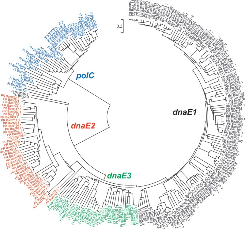
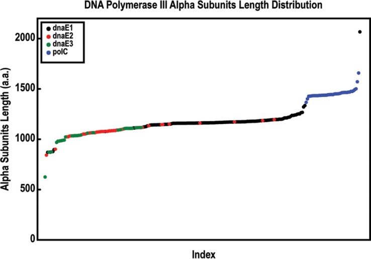
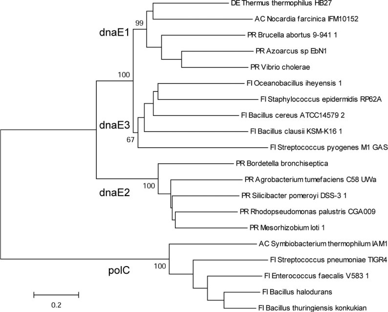
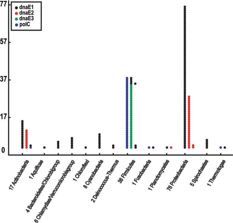
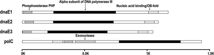
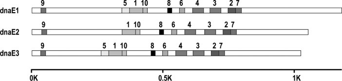
DNA polymerase III is one of the five eubacterial DNA polymerases that is responsible for the replication of DNA duplex. Among the ten subunits of the DNA polymerase III core enzyme, the alpha subunit catalyzes the reaction for polymerizing both DNA strands. In this study, we extracted genomic sequences of the alpha subunit from 159 sequenced eubacterial genomes, and carried out sequence-based phylogenetic and structural analyses. We found that all eubacterial genomes have one or more alpha subunits, which form either homodimers or heterodimers. Phylogenetic and domain structural analyses as well as copy number variations of the alpha subunit in each bacterium indicate the classification of alpha subunit into four basic groups: polC, dnaE1, dnaE2, and dnaE3. This classification is of essence in genome composition analysis. We also consolidated the naming convention to avoid further confusion in gene annotations.
Figures
Similar articles
-
Comprehensive analysis of DNA polymerase III α subunits and their homologs in bacterial genomes.Nucleic Acids Res. 2014 Feb;42(3):1393-413. doi: 10.1093/nar/gkt900. Epub 2013 Oct 7. Nucleic Acids Res. 2014. PMID: 24106089 Free PMC article.
-
GC content variability of eubacteria is governed by the pol III alpha subunit.Biochem Biophys Res Commun. 2007 Apr 27;356(1):20-5. doi: 10.1016/j.bbrc.2007.02.109. Epub 2007 Feb 28. Biochem Biophys Res Commun. 2007. PMID: 17336933
-
Nucleotide compositional asymmetry between the leading and lagging strands of eubacterial genomes.Res Microbiol. 2010 Dec;161(10):838-46. doi: 10.1016/j.resmic.2010.09.015. Epub 2010 Sep 22. Res Microbiol. 2010. PMID: 20868744
-
Bacterial replicases and related polymerases.Curr Opin Chem Biol. 2011 Oct;15(5):587-94. doi: 10.1016/j.cbpa.2011.07.018. Epub 2011 Aug 19. Curr Opin Chem Biol. 2011. PMID: 21855395 Free PMC article. Review.
-
Structure and function of eukaryotic DNA polymerase δ.Subcell Biochem. 2012;62:217-36. doi: 10.1007/978-94-007-4572-8_12. Subcell Biochem. 2012. PMID: 22918588 Review.
Cited by
-
Biophysical characterization of DNA binding from single molecule force measurements.Phys Life Rev. 2010 Sep;7(3):299-341. doi: 10.1016/j.plrev.2010.06.001. Epub 2010 Jun 4. Phys Life Rev. 2010. PMID: 20576476 Free PMC article. Review.
-
On the molecular mechanism of GC content variation among eubacterial genomes.Biol Direct. 2012 Jan 10;7:2. doi: 10.1186/1745-6150-7-2. Biol Direct. 2012. PMID: 22230424 Free PMC article.
-
Comprehensive analysis of DNA polymerase III α subunits and their homologs in bacterial genomes.Nucleic Acids Res. 2014 Feb;42(3):1393-413. doi: 10.1093/nar/gkt900. Epub 2013 Oct 7. Nucleic Acids Res. 2014. PMID: 24106089 Free PMC article.
-
DNA repair and genome maintenance in Bacillus subtilis.Microbiol Mol Biol Rev. 2012 Sep;76(3):530-64. doi: 10.1128/MMBR.05020-11. Microbiol Mol Biol Rev. 2012. PMID: 22933559 Free PMC article. Review.
-
The pendulum model for genome compositional dynamics: from the four nucleotides to the twenty amino acids.Genomics Proteomics Bioinformatics. 2012 Aug;10(4):175-80. doi: 10.1016/j.gpb.2012.08.002. Epub 2012 Aug 11. Genomics Proteomics Bioinformatics. 2012. PMID: 23084772 Free PMC article.
References
-
- Goodman M.F. Error-prone repair DNA polymerases in prokaryotes and eukaryotes. Annu. Rev. Biochem. 2002;71:17–50. - PubMed
-
- Lewin B. Oxford University Press; Oxford, UK: 2004. Genes VIII.
-
- Lehman I.R. Discovery of DNA polymerase. J. Biol. Chem. 1989;278:34733–34738. - PubMed
-
- Kobayashi S. Fidelity of Escherichia coli DNA polymerase IV. Preferential generation of small deletion mutations by dNTP-stabilized misalignment. J. Biol. Chem. 2002;277:34198–34207. - PubMed
Publication types
MeSH terms
Substances
LinkOut - more resources
Full Text Sources
Molecular Biology Databases
