Review
. 2007:79:589-620.
doi: 10.1016/S0074-7742(07)79026-8.

Remyelination in multiple sclerosis

Affiliations
Review

Remyelination in multiple sclerosis

Divya M Chari. Int Rev Neurobiol. 2007.

Abstract

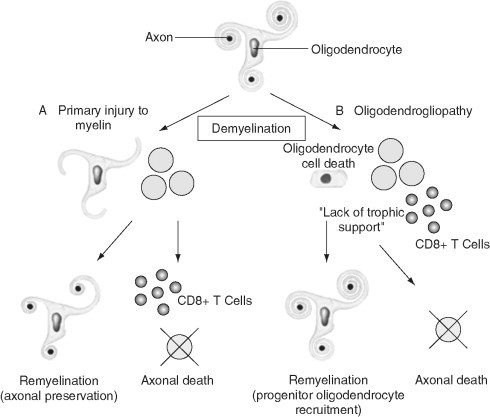
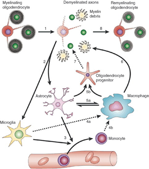
Remyelination is the phenomenon by which new myelin sheaths are generated around axons in the adult central nervous system (CNS). This follows the pathological loss of myelin in diseases like multiple sclerosis (MS). Remyelination can restore conduction properties to axons (thereby restoring neurological function) and is increasingly believed to exert a neuroprotective role on axons. Remyelination occurs in many MS lesions but becomes increasingly incomplete/inadequate and eventually fails in the majority of lesions and patients. Efforts to understand the causes for this failure of regeneration have fueled research into the biology of remyelination and the complex, interdependent cellular and molecular factors that regulate this process. Examination of the mechanisms of repair of experimental lesions has demonstrated that remyelination occurs in two major phases. The first consists of colonization of lesions by oligodendrocyte progenitor cells (OPCs), the second the differentiation of OPCs into myelinating oligodendrocytes that contact demyelinated axons to generate functional myelin sheaths. Several intracellular and extracellular molecules have been identified that mediate these two phases of repair. Theoretically, the repair of demyelinating lesions can be promoted by enhancing the intrinsic repair process (by providing one or more remyelination-enhancing factors or via immunoglobulin therapy). Alternatively, endogenous repair can be bypassed by introducing myelinogenic cells into demyelinated areas; several cellular candidates have been identified that can mediate repair of experimental demyelinating lesions. Future challenges confronting therapeutic strategies to enhance remyelination will involve the translation of findings from basic science to clinical demyelinating disease.


Figures

Fig. 1
Fig. 1
Section from the brain of an MS patient stained with Luxol fast blue to identify subcortical white matter (WM). Green arrows indicate three areas of myelin loss which represent foci of chronic demyelination. Red arrows indicate areas of pale myelin staining which represent the shadow plaques where remyelination has occurred. This illustrates the key point that spontaneous remyelination can occur efficiently in MS but that this process fails on many occasions, leaving axons chronically demyelinated. Reproduced with permission from Adams (1989).
Fig. 2
Fig. 2
There are two options following demyelination in multiple sclerosis: either (A) remyelination can take place with axonal preservation or (B) axonal death may ensue, either due to a range of cytotoxicity mechanisms or due to failure of local neurotrophic support due to glial cell injury. Remyelination most likely occurs by the recruitment of OPCs into demyelinated areas that subsequently mature into myelinating cells. Modified with permission from Rodriguez (2003).
Fig. 3
Fig. 3
OPCs increase expression of Nkx2.2 mRNA and Olig2 mRNA in response to CNS demyelination. (A) Diagram to show focal demyelinating lesions in the caudal cerebellar peduncle (CCP), a large WM tract in adult rats, created by stereotaxic injection of ethidium bromide (EB). (B) This creates focally demyelinated areas, identifiable by absence of myelin basic protein (MBP) mRNA following in situ hybridization. (C) Toluidine blue‐stained resin section at 5 days postlesion showing demyelinated axons in transverse section and debris‐filled macrophages. (D) All demyelinated axons have been reinvested with thin myelin sheaths that are characteristic of CNS remyelination, at 4 weeks postlesion. (E–H) PDGFRα, Nkx2.2, Olig1, and Olig2 mRNA expression in normal WM detected by in situ hybridization. (J–M) The expression of the same mRNAs in areas of demyelination 5 days postlesion. Scale bar = 1.5 mm (A), 25 μm (C–D), and 100 μm (E–N). Modified with permission from Zhao et al. (2005).
Fig. 4
Fig. 4
Chronic MS lesions contain O4‐positive OPCs that fail to bind the Ki67 antibody (a marker of cell proliferation), suggesting that these are a relatively quiescent population. Some lesions do contain some Ki67‐immunoreactive nuclei, such as the one indicated toward the lower right of the figure. Reproduced with permission from Wolswijk (1998).
Fig. 5
Fig. 5
Corticosteroid (CS) treatment significantly reduces oligodendrocyte‐mediated remyelination of EB‐induced lesions in the spinal cord at 1 month. Treatment groups consisted of injection of high‐dose MPS with and without subsequent PRED taper, in order to mimic current CS treatment regimens used clinically to treat acute relapses in MS. (A) Extensive oligodendrocyte and Schwann cell‐mediated remyelination in saline‐injected control rats. (B, C) Reduced oligodendrocyte remyelination in CS‐treated rats with Schwann cell remyelination unaffected. Oligodendrocyte remyelination is indicated with arrows. MPS, methylprednisolone, PRED, prednisone. Modified with permission from Chari et al. (2006a).
Fig. 6
Fig. 6
Diagram illustrating that remyelination involves a complex sequence of tightly coordinated events, the “dysregulation” of which will result in remyelination impairment. In response to a demyelinating insult, the myelinated axons undergo demyelination (1). This generates myelin debris. Demyelination causes activation of resident astrocytes and microglia (2). Activated astrocytes and microglia produce factors that act to recruit monocytes from the vasculature (3). Microglia (4a) and recruited monocytes (4b) differentiate into macrophages. Activated astrocytes and macrophages produce factors that can mutually activate each other (5a). As a result of such activation, both produce growth factors that act on OPCs to alter their proliferation and differentiation (5b). Macrophages remove myelin debris (6), a function that is beneficial for remyelination. Under the influence of factors produced by astrocytes and macrophages, recruited OPCs engage with demyelinated axons (7) and differentiate into remyelinating oligodendrocytes (8). Modified with permission from Franklin (2002).
Fig. 7
Fig. 7
Transplantation of human ESC‐derived predifferentiated oligodendrocyte progenitors into a region of demyelination results in differentiation of ESCs into oligodendrocytes and remyelination. (A) Assessment of cell fate and differentiation following transplantation using double immunohistochemical staining for human nuclei (green) and the oligodendrocyte marker APC‐CC1 (red) confirms that transplanted cells survive and differentiate into mature oligodendrocytes. (B) Electron micrograph (EM) of the transplant environment 7 days postinjury. Asterisks indicate demyelinated axons in an astrogliosis‐free microenvironment. (C) EM of a rat spinal cord that was transplanted with human ESC‐derived oligodendrocyte progenitors 7 days postinjury. Oligodendrocyte‐remyelinated axons (R; with characteristically thin myelin sheaths) are evident among few normally myelinated axons (N). (D) EM of a spinal cord injury site 10 months postinjury, illustrating an astrocyte (A) with a large intermediate filament‐rich process (AP) extending to demyelinated axons (arrows) and myelinated axons (arrowheads), both surrounded by intermediate filament‐rich astrocytic processes. This illustrates the point that demyelinated axons in areas of chronic injury are ensheathed by astrocytic processes, which may prevent remyelination by endogenous or transplanted cells. Reproduced with permission from Keirstead (2005).

References

    1. Adams C.W.M. “A Colour Atlas of Multiple Sclerosis and Other Myelin Disorders.”. Wolfe Medical Publications; London: 1989.
    1. Antony J.M., van Marle G., Opii W., Butterfield D.A., Mallet F., Yong V.W., Wallace J.L., Deacon R.M., Warren K., Power C. Human endogenous retrovirus glycoprotein‐mediated induction of redox reactants causes oligodendrocyte death and demyelination. Nat. Neurosci. 2004;7:1088–1095. - PubMed
    1. Arnett H.A., Mason J., Marino M., Suzuki K., Matsushima G.K., Ting J.P. TNF alpha promotes proliferation of oligodendrocyte progenitors and remyelination. Nat. Neurosci. 2001;4:1116–1122. - PubMed
    1. Arnett H.A., Wang Y., Matsushima G.K., Suzuki K., Ting J.P. Functional genomic analysis of remyelination reveals importance of inflammation in oligodendrocyte regeneration. J. Neurosci. 2003;23:9824–9832. - PMC - PubMed
    1. Arnett H.A., Fancy S.P., Alberta J.A., Zhao C., Plant S.R., Kaing S., Raine C.S., Rowitch D.H., Franklin R.J., Stiles C.D. bHLH transcription factor Olig1 is required to repair demyelinated lesions in the CNS. Science. 2004;17:2111–2115. - PubMed

Publication types