

Calsequestrin mutation and catecholaminergic polymorphic ventricular tachycardia: a simulation study of cellular mechanism
- PMID: 17531962
- PMCID: PMC2030636
- DOI: 10.1016/j.cardiores.2007.04.010
Calsequestrin mutation and catecholaminergic polymorphic ventricular tachycardia: a simulation study of cellular mechanism
Abstract
Objectives: Patients with a missense mutation of the calsequestrin 2 gene (CASQ2) are at risk for catecholaminergic polymorphic ventricular tachycardia. This mutation (CASQ2(D307H)) results in decreased ability of CASQ2 to bind Ca2+ in the sarcoplasmic reticulum (SR). In this theoretical study, we investigate a potential mechanism by which CASQ2(D307H) manifests its pro-arrhythmic consequences in patients.
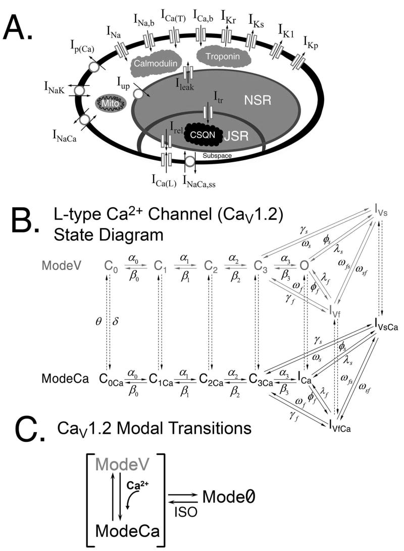
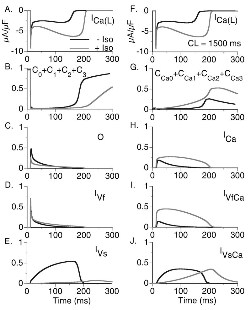
Methods: Using simulations in a model of the guinea pig ventricular myocyte, we investigate the mutation's effect on SR Ca2+ storage, the Ca2+ transient (CaT), and its indirect effect on ionic currents and membrane potential. We model the effects of isoproterenol (ISO) on Ca(V)1.2 (the L-type Ca2+ current, I(Ca(L))) and other targets of beta-adrenergic stimulation.
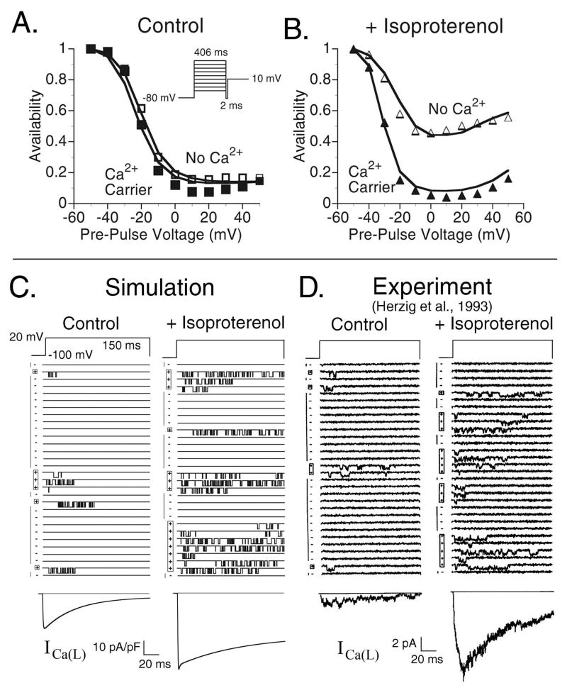
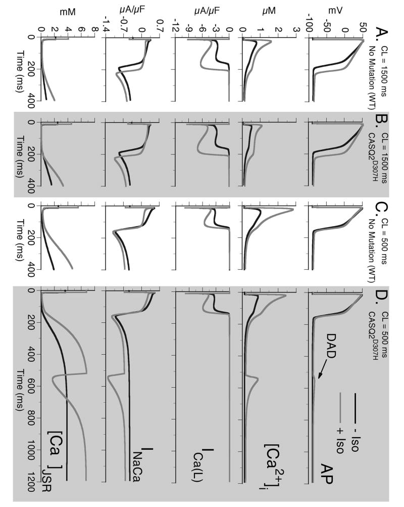
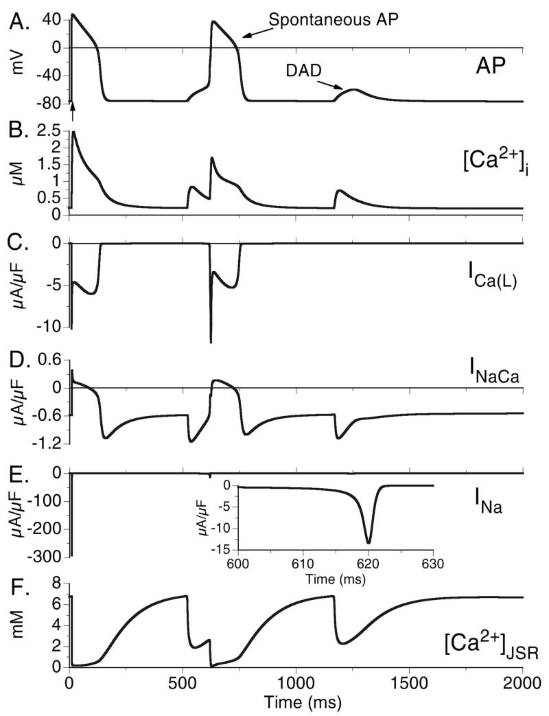
Results: ISO increases I(Ca(L)), prolonging action potential (AP) duration (Control: 172 ms, +ISO: 207 ms, at cycle length of 1500 ms) and increasing CaT (Control: 0.79 microM, +ISO: 1.61 microM). ISO increases I(Ca(L)) by reducing the fraction of channels which undergo voltage-dependent inactivation and increasing transitions from a non-conducting to conducting mode of channel gating. CASQ2(D307H) reduces SR storage capacity, thereby reducing the magnitude of CaT (Control: 0.79 microM, CASQ2(D307H): 0.52 microM, at cycle length of 1500 ms). The combined effect of CASQ2(D307H) and ISO elevates SR free Ca2+ at a rapid rate, leading to store-overload-induced Ca2+ release and delayed afterdepolarization (DAD). If resting membrane potential is sufficiently elevated, the Na+-Ca2+ exchange-driven DAD can trigger I(Na) and I(Ca(L)) activation, generating a triggered arrhythmogenic AP.
Conclusions: The CASQ2(D307H) mutation manifests its pro-arrhythmic consequences due to store-overload-induced Ca2+ release and DAD formation due to excess free SR Ca2+ following rapid pacing and beta-adrenergic stimulation.
Figures
Comment in
-
Sarcoplasmic reticulum Ca2+ release channel complex and automatism: a matter of fine tuning.Cardiovasc Res. 2007 Jul 1;75(1):7-9. doi: 10.1016/j.cardiores.2007.04.029. Epub 2007 May 3. Cardiovasc Res. 2007. PMID: 17524377 No abstract available.
Similar articles
-
Abnormal calcium signaling and sudden cardiac death associated with mutation of calsequestrin.Circ Res. 2004 Mar 5;94(4):471-7. doi: 10.1161/01.RES.0000115944.10681.EB. Epub 2004 Jan 8. Circ Res. 2004. PMID: 14715535
-
Functional consequences of stably expressing a mutant calsequestrin (CASQ2D307H) in the CASQ2 null background.Am J Physiol Heart Circ Physiol. 2012 Jan 1;302(1):H253-61. doi: 10.1152/ajpheart.00578.2011. Epub 2011 Oct 7. Am J Physiol Heart Circ Physiol. 2012. PMID: 21984545 Free PMC article.
-
A mutation in calsequestrin, CASQ2D307H, impairs Sarcoplasmic Reticulum Ca2+ handling and causes complex ventricular arrhythmias in mice.Cardiovasc Res. 2007 Jul 1;75(1):69-78. doi: 10.1016/j.cardiores.2007.03.002. Epub 2007 Mar 12. Cardiovasc Res. 2007. PMID: 17449018 Free PMC article.
-
Calsequestrin mutations and catecholaminergic polymorphic ventricular tachycardia.Pediatr Cardiol. 2012 Aug;33(6):959-67. doi: 10.1007/s00246-012-0256-1. Epub 2012 Mar 16. Pediatr Cardiol. 2012. PMID: 22421959 Free PMC article. Review.
-
Calsequestrin 2 and arrhythmias.Am J Physiol Heart Circ Physiol. 2012 Mar 15;302(6):H1250-60. doi: 10.1152/ajpheart.00779.2011. Epub 2011 Dec 23. Am J Physiol Heart Circ Physiol. 2012. PMID: 22198169 Free PMC article. Review.
Cited by
-
Calcium/calmodulin-dependent protein kinase II (CaMKII) inhibition ameliorates arrhythmias elicited by junctin ablation under stress conditions.Heart Rhythm. 2015 Jul;12(7):1599-610. doi: 10.1016/j.hrthm.2015.03.043. Epub 2015 Mar 23. Heart Rhythm. 2015. PMID: 25814413 Free PMC article.
-
Ionic mechanisms of arrhythmogenesis.Trends Cardiovasc Med. 2015 Aug;25(6):487-96. doi: 10.1016/j.tcm.2015.01.005. Epub 2015 Jan 16. Trends Cardiovasc Med. 2015. PMID: 25701094 Free PMC article. Review.
-
Delayed afterdepolarization-induced triggered activity in cardiac purkinje cells mediated through cytosolic calcium diffusion waves.Physiol Rep. 2019 Dec;7(24):e14296. doi: 10.14814/phy2.14296. Physiol Rep. 2019. PMID: 31872561 Free PMC article.
-
Electrodiffusion dynamics in the cardiomyocyte dyad at nano-scale resolution using the Poisson-Nernst-Planck (PNP) equations.PLoS Comput Biol. 2025 Jun 12;21(6):e1013149. doi: 10.1371/journal.pcbi.1013149. eCollection 2025 Jun. PLoS Comput Biol. 2025. PMID: 40505081 Free PMC article.
-
Cardiovascular disease models: A game changing paradigm in drug discovery and screening.Biomaterials. 2019 Apr;198:3-26. doi: 10.1016/j.biomaterials.2018.09.036. Epub 2018 Oct 1. Biomaterials. 2019. PMID: 30343824 Free PMC article. Review.
References
-
- Beard NA, Laver DR, Dulhunty AF. Calsequestrin and the calcium release channel of skeletal and cardiac muscle. Prog Biophys Mol Biol. 2004;85:33–69. - PubMed
-
- Lahat H, Pras E, Olender T, Avidan N, Ben-Asher E, Man O, et al. A missense mutation in a highly conserved region of CASQ2 is associated with autosomal recessive catecholamine-induced polymorphic ventricular tachycardia in Bedouin families from Israel. Am J Hum Genet. 2001;69:1378–84. - PMC - PubMed
-
- Viatchenko-Karpinski S, Terentyev D, Györke I, Terentyeva R, Volpe P, Priori SG, et al. Abnormal calcium signaling and sudden cardiac death associated with mutation of calsequestrin. Circ Res. 2004;94:471–7. - PubMed
-
- Houle TD, Ram ML, Cala SE. Calsequestrin mutant D307H exhibits depressed binding to its protein targets and a depressed response to calcium. Cardiovasc Res. 2004;64:227–33. - PubMed
Publication types
MeSH terms
Substances
Grants and funding
LinkOut - more resources
Full Text Sources
Research Materials
Miscellaneous
