. 2007 Sep 30;366(2):293-303.
doi: 10.1016/j.virol.2007.04.029. Epub 2007 May 25.

The nonstructural protein 8 (nsp8) of the SARS coronavirus interacts with its ORF6 accessory protein

Affiliations

The nonstructural protein 8 (nsp8) of the SARS coronavirus interacts with its ORF6 accessory protein

Purnima Kumar et al. Virology. .

Abstract

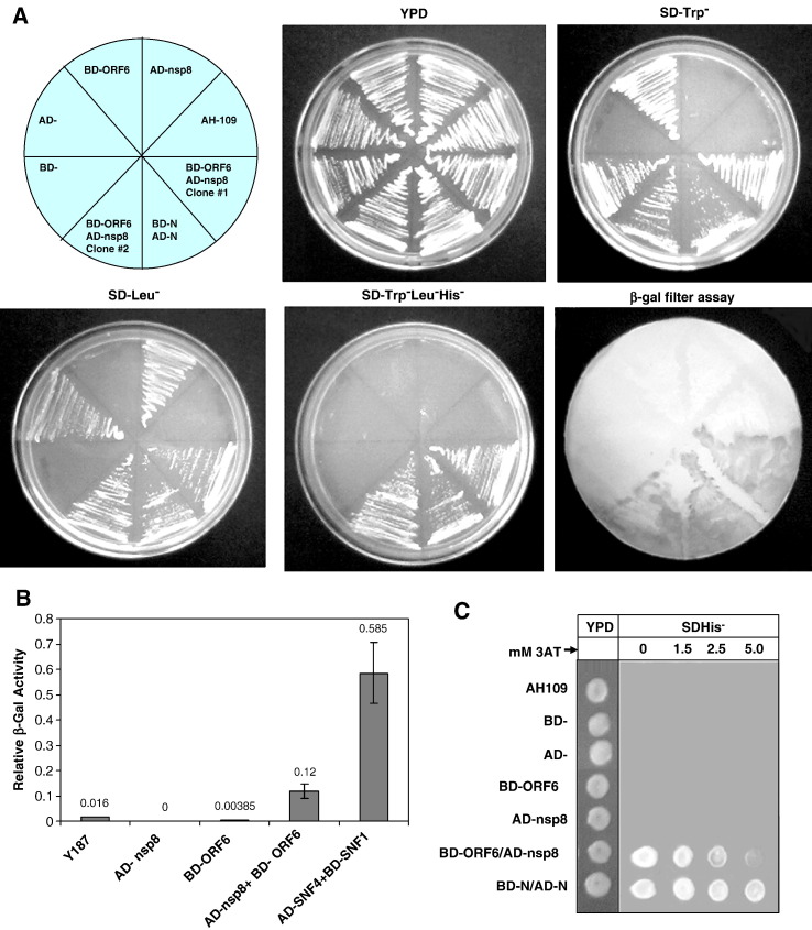
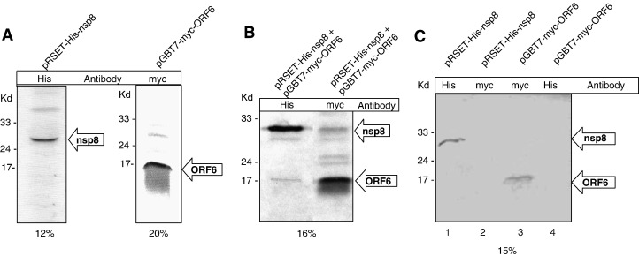
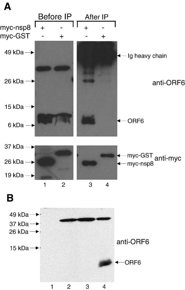
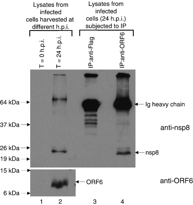
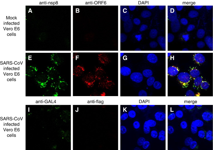
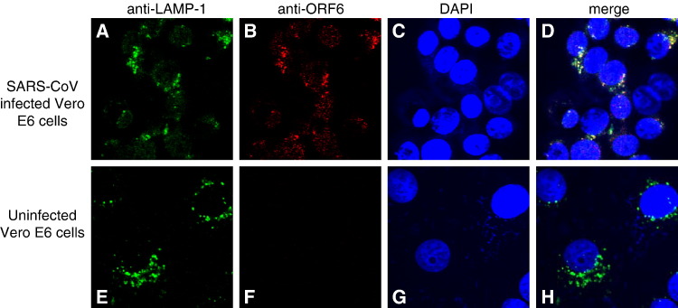
Severe acute respiratory syndrome (SARS) coronavirus (SARS-CoV) caused a severe outbreak in several regions of the world in 2003. The SARS-CoV genome is predicted to contain 14 functional open reading frames (ORFs). The first ORF (1a and 1b) encodes a large polyprotein that is cleaved into nonstructural proteins (nsp). The other ORFs encode for four structural proteins (spike, membrane, nucleocapsid and envelope) as well as eight SARS-CoV-specific accessory proteins (3a, 3b, 6, 7a, 7b, 8a, 8b and 9b). In this report we have cloned the predicted nsp8 gene and the ORF6 gene of the SARS-CoV and studied their abilities to interact with each other. We expressed the two proteins as fusion proteins in the yeast two-hybrid system to demonstrate protein-protein interactions and tested the same using a yeast genetic cross. Further the strength of the interaction was measured by challenging growth of the positive interaction clones on increasing gradients of 2-amino trizole. The interaction was then verified by expressing both proteins separately in-vitro in a coupled-transcription translation system and by coimmunoprecipitation in mammalian cells. Finally, colocalization experiments were performed in SARS-CoV infected Vero E6 mammalian cells to confirm the nsp8-ORF6 interaction. To the best of our knowledge, this is the first report of the interaction between a SARS-CoV accessory protein and nsp8 and our findings suggest that ORF6 protein may play a role in virus replication.


Figures

Fig. 1
Fig. 1
Cloning of the putative nsp8 and ORF6 genes from the SARS CoV genome. The full-length nsp8 (198 amino acids) and ORF6 (66 amino acids) genes of the SARS coronavirus (Tor2 isolate) were PCR-amplified from a genomic construct of clone NC_004718 and cloned into the pCR-XL-TOPO vector (Invitrogen) as described in Table 1. The scale on top shows genomic region.
Fig. 2
Fig. 2
Yeast two-hybrid results showing that nsp8 and ORF6 proteins interact with each other. (A) AD-nsp8 fusion protein and BD-ORF6 fusion protein tested as single and co-transformants on various synthetic growth media lacking specific amino-acids. YPD media shows uninhibited growth of all transformants and host strain. SDTrp and SD-Leu selective media have Tryptophan and Leucine missing, respectively. SD TrpLeuHis media are triple dropout plates lacking Tryptophan, Leucine and Histidine. Growth on the SD TrpLeuHis plate and blue color (shown here in shades of grey) on the β-gal filter assay represents a positive interaction. BD-N:AD-N combination was used as a positive control in these yeast two-hybrid experiments (Surjit et al., 2004). (B) Liquid β-galactosidase assay results of the nsp8–ORF6 protein–protein interactions. Single transformants and co-transformants were analyzed in a liquid β-galactosidase assay and compared with each other. Values given are in arbitrary units. The numbers above each bar represent the mean from five independent transformants. Y187 corresponds to the untransformed host strain. Transformants with more than one plasmid are separated by a slash. Positive controls used in this assay are denoted by BD-SNF1/AD-SNF4 (Harper et al., 1993). (C) Measurement of strength of the nsp8–ORF6 interactions on an increasing 3-AT gradient. Activation of the HIS3 reporter was determined for the host AH109 strain, single transformants (BD, AD, BD-ORF6 and AD-nsp8), and co-transformants (BD-ORF6/AD-nsp8 and BD-N/AD-N). Hundred-fold serial dilutions of all of the above-mentioned log-phase cultures were plated on YPD (left) followed by SD-His-plus 50 mM 3-AT in increasing concentrations (0 mM to 5 mM 3-AT).
Fig. 3
Fig. 3
Confirmation of the nsp8–ORF6 interaction using an in-vitro cell-free coupled transcription/translation coimmunoprecipitation assay. (A) The His6-tagged nsp8 and myc-tagged ORF6 proteins were produced by coupled transcription–translation in the presence of 35S-Met and detected using their corresponding antibodies (Anti-His and anti-myc, respectively). Both nsp8 and ORF6 proteins were visible and corresponded to their correct molecular sizes on 12% and 20% SDS-PAGE, respectively. (B) The 35S-Met-His-nsp8 and 35S-Met-myc-ORF6 proteins were detected by autoradiography. The nsp8–ORF6 complex was detected by both anti-His and anti-myc antibodies on 16% SDS-PAGE. When nsp8 was probed for using anti-His antibody, the ORF6 protein was visible and when ORF6 protein was probed using myc antibody, the nsp8 protein was visible. (C) In a control reaction, the 35S-Met-His6-nsp8 and 35S-Met-myc-ORF6 proteins showed no cross-reactivity with the other antibody used in the pull-down experiments. Also this control experiment shows that the proteins do not non-specifically bind to the beads used in the results shown in panel B. Lane 1 shows the nsp8 protein immunoprecipitated by anti-His, lane 2 does not show nsp8 protein band when immunoprecipitated by anti-myc. Similarly, lane 3 shows myc-tagged ORF6 protein immunoprecipitated by anti-myc and lane 4 does not show ORF6 protein where it was immunoprecipitated using anti-His.
Fig. 4
Fig. 4
(A) Cell lysates were obtained from Vero E6 cells expressing ORF6 and myc-nsp8 or ORF6 and myc-GST (negative control) and subjected to Western blot analysis with either anti-ORF6 polyclonal (upper panel, lanes 1 and 2) or anti-myc monoclonal antibodies (lower panel, lanes 1 and 2). Equal amounts of lysates were subjected to immunoprecipitation using anti-myc polyclonal antibodies and protein A beads. The immuno-complexes were subjected to Western blot with either anti-ORF6 polyclonal (upper panel, lanes 3 and 4) or anti-myc monoclonal antibodies (lower panel, lanes 3 and 4). (B) Equal amounts of total cellular protein from Vero E6 cells either uninfected (lane 1) or infected with wildtype Dryvax (lane 2), a recombinant vaccinia virus expressing the SARS-CoV S protein (lane 3), or the recombinant vaccinia virus expressing the ORF6 protein used in this study (lane 4) were subjected to Western blot and probed with anti-ORF6 rabbit polyclonal antibody. This demonstrates that the ∼ 37 kDa band seen in lanes 1 and 2 of panel A is a cross-reacting protein that is probably a vaccinia virus protein or an up-regulated cellular protein which is detected by the rabbit anti-ORF6 antibody used in this study.
Fig. 5
Fig. 5
Lysates were obtained from SARS-CoV infected Vero E6 cells harvested at 0 or 24 h post infection (h.p.i.) and Western blot analyses were performed to determine the expression of SARS-CoV proteins using mouse anti-nsp8 monoclonal antibody (upper panel, lanes 1 and 2) and rabbit anti-ORF6 polyclonal antibody (lower panel, lanes 1 and 2). Cell lysates obtained at 24 h.p.i. were then subjected to immunoprecipitation using anti-Flag (irrelevant antibody as negative control) or anti-ORF6 rabbit polyclonal antibodies and protein A beads. The immuno-complexes were subjected to Western blot analysis with mouse anti-nsp8 monoclonal antibody (upper panel, lanes 3 and 4).
Fig. 6
Fig. 6
Indirect immunofluorescence used to study cellular localization of nsp8 and ORF6 in SARS-CoV infected Vero E6 cells. (Upper panel) Mouse anti-nsp8 (A) and rabbit anti-ORF6 (B) antibodies were used against uninfected cells to determine if the antibodies showed any unspecific staining. Nuclei are represented by means of a DAPI stain (C). (Middle panel) Vero E6 cells infected with SARS-CoV were examined by indirect immunofluorescence using the same antibodies. The expression of nsp8 is represented by FITC staining (E), while the expression of ORF6 is represented by Rhodamine staining (F). Nuclei are represented by means of a DAPI stain (G). The merged image showed that the nsp8 and ORF6 proteins colocalize to punctuate structures in the cytoplasm (H). (Lower panel) Irrelevant antibodies (mouse anti-GAL4TA (I) and rabbit anti-flag (J)) were used to probe SARS-CoV infected cells at the same final concentrations as their counterparts in the middle panel to show the specificity of the antibodies used in detecting the ORF6 and nsp8 proteins.
Fig. 7
Fig. 7
Indirect immunofluorescence was used to probe for LAMP-1 (A, E) and ORF6 (B, F) staining in SARS-CoV infected cells (upper panel) and uninfected Vero E6 cells (lower panel). Nuclei are represented by means of a DAPI stain (C, G). The merged image showed that there is colocalization between the LAMP-1 protein and ORF6 proteins in SARS-CoV infected cells (D, upper panel).

Similar articles

Cited by

References

    1. Denison M.R., Zoltick P.W., Hughes S.A., Giangreco B., Olson A.L., Perlman S., Leibowitz J.L., Weiss S.R. Intracellular processing of the N-terminal ORF 1a proteins of the coronavirus MHV-A59 requires multiple proteolytic events. Virology. 1992;189(1):274–284. - PMC - PubMed
    1. Fielding B.C., Tan Y.J., Shuo S., Tan T.H., Ooi E.E., Lim S.G., Hong W., Goh P.Y. Characterization of a unique group-specific protein (U122) of the severe acute respiratory syndrome coronavirus. J. Virol. 2004;78(14):7311–7318. - PMC - PubMed
    1. Geng H., Liu Y.M., Chan W.S., Lo A.W., Au D.M., Waye M.M., Ho Y.Y. The putative protein 6 of the severe acute respiratory syndrome-associated coronavirus: expression and functional characterization. FEBS Lett. 2005;579(30):6763–6768. - PMC - PubMed
    1. Harper J.W., Adami G.R., Wei N., Keyomarsi K., Elledge S.J. The p21 Cdk-interacting protein Cip1 is a potent inhibitor of G1 cyclin-dependent kinases. Cell. 1993;75(4):805–816. - PubMed
    1. Holzer G.W., Falkner F.G. Construction of a vaccinia virus deficient in the essential DNA repair enzyme uracil DNA glycosylase by a complementing cell line. J. Virol. 1997;71(7):4997–5002. - PMC - PubMed

Publication types

Substances