. 2007 Sep;128(1-2):1-8.
doi: 10.1016/j.virusres.2007.02.007. Epub 2007 May 25.

Up-regulation of IL-6 and TNF-alpha induced by SARS-coronavirus spike protein in murine macrophages via NF-kappaB pathway

Affiliations

Up-regulation of IL-6 and TNF-alpha induced by SARS-coronavirus spike protein in murine macrophages via NF-kappaB pathway

Wei Wang et al. Virus Res. 2007 Sep.

Abstract

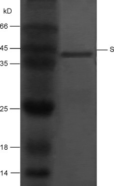
The clinical picture of severe acute respiratory syndrome (SARS) is characterized by an over-exuberant immune response with lung lymphomononuclear cells infilteration and proliferation that may account for tissue damage more than the direct effect of viral replication. To understand how cells response in the early stage of virus-host cell interaction, in this study, a purified recombinant S protein was studied for stimulating murine macrophages (RAW264.7) to produce proinflammatory cytokines (IL-6 and TNF-alpha) and chemokine IL-8. We found that direct induction of IL-6 and TNF-alpha release in the supernatant in a dose-, time-dependent manner and highly spike protein-specific, but no induction of IL-8 was detected. Further experiments showed that IL-6 and TNF-alpha production were dependent on NF-kappaB, which was activated through I-kappaBalpha degradation. These results suggest that SARS-CoV spike protein may play an important role in the pathogenesis of SARS, especially in inflammation and high fever.


Figures

Fig. 1
Fig. 1
Expression and purification of recombinant S protein of SARS-CoV. Recombinant S protein was expressed in E. coli after 5 h induction with IPTG, purified by Ni-NTA affinity column, separated by 12% SDS-PAGE and visualized by Coomassie Brilliant Blue staining.
Fig. 2
Fig. 2
Purified S protein of SARS-CoV promotes IL-6 and TNF-α induction in murine macrophages. IL-6 and TNF-α were induced by S protein in a dose-dependent manner. RAW264.7 cells were incubated with the indicated concentrations of S protein for 12 h, IL-6 and TNF-α (A and B) production were determined by ELISA. IL-6 and TNF-α were induced by S protein in a time-dependent manner. RAW264.7 cells were incubated with 20 μg/ml S protein for the indicated times, IL-6 and TNF-α (C and D) production were determined by ELISA. (E) Transciptional induction of IL-6 by S protein. RAW264.7 cells were incubated with the indicated concentrations of S protein for 12 h, and the mRNA levels of IL-6 were tested by RT-PCR. Data shown are representative of three independent experiments. IL-6 and TNF-α were induced in an S protein-specific manner. RAW264.7 cells were untreated, incubated with 20 μg/ml S protein, 20 μg/ml S protein pre-treated by Detoxi-GelTM Endotoxin Removing Gel, 20 μg/ml S protein pre-boiled for 1 h or pre-incubated with anti-S antibody for 2 h at 37 °C, 20 μg/ml GFP and 20 μg/ml BSA. IL-6 and TNF-α (F and G) production were determined by ELISA. All bars represent the means ± S.E. of three independent experiments.
Fig. 3
Fig. 3
NF-κB activation by purified S protein of SARS-CoV. (A) RAW264.7 cells in a 96-well plate were transfected with NF-κB-luciferase reporter plasmid (0.1 μg/well). A Renila reporter plasmid (5 ng/well) was cotransfected for normalization. Twenty-four hour after transfection, the cells were either treated with S protein (10 μg/ml) or left untreated. After 12 h, the cells were lysed, and luciferase activity was quantified. The results were expressed as the mean ± S.E. of three independent experiments. (B) RAW264.7 cells in a 6-well plate were treated with S protein (10 μg/ml) or left untreated for 1.5 h. Total cell lysates were analyzed by Western blotting using anti-I-κBα polyclonal antibodies (upper panel) and anti-actin polyclonal antibodies (lower panel). The density of the protein band was determined by using Bio-Rad Quantity One imaging software. The values in parentheses are density values of I-κBα relative to actin. Results of one of three independent experiments are shown.
Fig. 4
Fig. 4
IL-6 and TNF-α induction by purified S protein of SARS-CoV requires NF-κB. RAW264.7 cells in a 24-well plate were transfected with either pcDNA3.1(+) (empty vector) or pcDNA3.1(+)-dn-NIK (expression plasmid for dominant negative mutant NIK). At 24 h posttransfection, the cells were treated with S protein (10 μg/ml) for 12 h, IL-6 and TNF-α (A and B) production in the supernatant were determined by ELISA. The means and standard error of the means are shown from three independent experiments.

Similar articles

Cited by

References

    1. Banerjee S., Narayanan K., Mizutani T., Makino S. Murine coronavirus replication-induced p38 mitogen-activated protein kinase activation promotes interleukin-6 production and virus replication in cultured cells. J. Virol. 2002;76(12):5937–5948. - PMC - PubMed
    1. Bieback K., Lien E., Klagge I.M., Avota E., Schneider-Schaulies J., Duprex W.P., Wagner H., Kirschning C.J., Ter Meulen V., Schneider-Schaulies S. Hemagglutinin protein of wild-type measles virus activates toll-like receptor 2 signaling. J. Virol. 2002;76(17):8729–8736. - PMC - PubMed
    1. Boehme K.W., Compton T. Innate sensing of viruses by toll-like receptors. J. Virol. 2004;78(15):7867–7873. - PMC - PubMed
    1. Chang Y.J., Liu C.Y., Chiang B.L., Chao Y.C., Chen C.C. Induction of IL-8 release in lung cells via activator protein-1 by recombinant baculovirus displaying severe acute respiratory syndrome-coronavirus spike proteins: identification of two functional regions. J. Immunol. 2004;173(12):7602–7614. - PubMed
    1. Chen Z., Zhang L., Qin C., Ba L., Yi C.E., Zhang F., Wei Q., He T., Yu W., Yu J., Gao H., Tu X., Gettie A., Farzan M., Yuen K.Y., Ho D.D. Recombinant modified vaccinia virus Ankara expressing the spike glycoprotein of severe acute respiratory syndrome coronavirus induces protective neutralizing antibodies primarily targeting the receptor binding region. J. Virol. 2005;79(5):2678–2688. - PMC - PubMed

MeSH terms