. 2007 Aug;41(2):188-96.
doi: 10.1016/j.bone.2007.04.183. Epub 2007 Apr 25.

Masticatory mechanics of a mandibular distraction osteogenesis site: interfragmentary micromovement

Affiliations

Masticatory mechanics of a mandibular distraction osteogenesis site: interfragmentary micromovement

Zongyang Sun et al. Bone. 2007 Aug.

Abstract

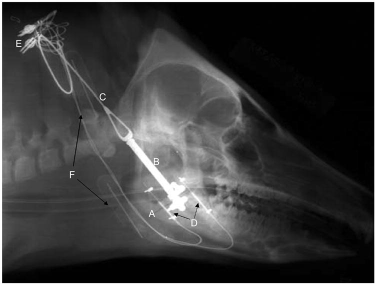
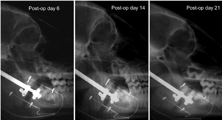
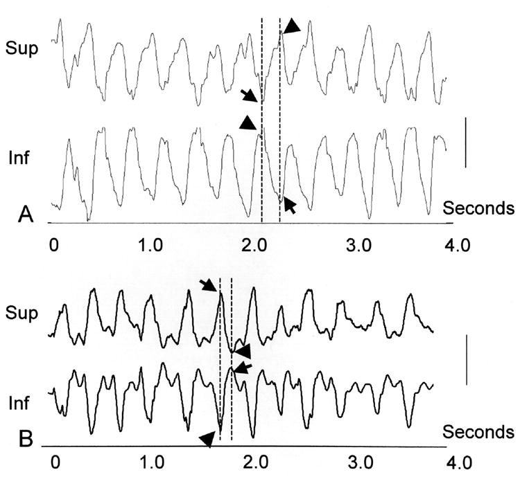
Micromovement at a fracture or distraction osteogenesis (DO) site may play a significant role in bone formation and healing. Mastication is an important physiological process that can cause substantial micromovement at a mandibular disjunction. The purpose of this study is to characterize and quantify the micromovement caused by mastication. Eighteen pigs, divided into three groups based on duration of consolidation, received a unilateral (right) mandibular angle distraction osteogenesis protocol. Differential variable reluctance transducers (DVRTs) and ultrasound crystals were used to measure the change of gap width as well as interfragmentary movement during mastication. Synchronized chewing video and interfragmentary movement recordings were used to determine the magnitude and direction of micromovement at different phases of the chewing cycle. The magnitude of micromovement did not increase significantly with distraction up to almost 5 mm, but did decrease gradually with consolidation. The average micromovement magnitude during the distraction phase was 0.2-0.3 mm, equaling 50,000-250,000 microstrain (muepsilon) on interfragmentary tissue. The dominant deformation pattern was bending in the sagittal plane. The most common direction of bending at the power stroke of chewing was concave dorsally, i.e., superior shortening and inferior lengthening. These findings elucidate how masticatory mechanics affect a mandibular distraction site, and the measurements may be useful for future simulation studies.


Figures

Fig. 1
Fig. 1
DO surgery and device placement. Post-operative lateral-view x-ray showing an osteotomy (A) at the mandibular angle. A distractor (B) was placed perpendicular to the osteotomy with an elongated handle (C) exiting posterior to the condylar neck. Measuring devices (D, DVRTs) were placed parallel to the distractor at superior and inferior locations. (E) The DVRT leads exited at the back of the neck. Antibiotic osmotic pumps (F) were inserted near the distractor handle and device wires for infection control.
Fig. 2
Fig. 2
Micromovement magnitude measurement. A typical crystal recording showing 12 chewing cycles in about 4.5 seconds was selected for analysis. The magnitude of micromovement was measured as the inter-peak distance (between broken lines), All cycles were measured and averaged. Calibration bar, 0.2 mm.
Fig. 3
Fig. 3
Serial x-rays of a distracted mandible (#C4). Note the uneven opening of the gap at the end of distraction. Pronounced new bone formation was seen after one week of consolidation (post-op day 14) and continued at two weeks of consolidation (post-op day 21), leading to decreased distraction space.
Fig. 4
Fig. 4
Examples of daily measurements of gap width change with time as measured by crystals or DVRTs. In successfully distracted animals, gap width increases linearly during the distraction phase (postoperative days 1-6). During the consolidation phase, gap width tends to decrease at the inferior location.
Fig. 5
Fig. 5
Micromovement directionality during mastication. Typical recordings from ultrasound crystals (A, pig #B4) and DVRTs (B, pig #C5), both exhibiting cyclic features. DVRT analog signals show the opposite direction of the true movement, i.e. a downward peak is lengthening while an upward peak is shortening. Two major peaks (broken lines) are present for each chewing cycle. The peak values (shortening, arrow; lengthening, arrow head) are opposite between the superior (Sup) and inferior (Inf) locations. Calibration bar, 0.2 mm.
Fig. 6
Fig. 6
A) Overview of a masticatory sequence showing micromovement (recorded by DVRTs) at superior (blue) and inferior (red) locations superimposed on the video. The vertical scanning lines (arrows) show the moment recorded. B) A single chewing cycle consisting of 23 frames (1/60 sec each). The selected frames (numbered) indicate the closing stroke (frames 1-7), the power stroke (frames 8-14) and the opening stroke (frames 15-23). During the power stroke, the superior location shortened, while the inferior location lengthened. During opening, these changes were reversed.
Fig. 7
Fig. 7
Masticatory micromovement magnitude over time (average and standard deviation). The “distracted” are those with at least 3 mm of distraction (n=15), while the “undistracted” group consists of 3 pigs with broken appliances and maximum distraction of 1.5 mm. Numbers beside each average value indicate the sample size that had available data. Vertical bars indicate standard deviations (upward, distracted group; downward, undistracted group). Note both superior and inferior locations showed a minor gradual increase during the distraction phase (days 1-6) and a more substantial decrease during the consolidation phase (days 7-21).
Fig. 8
Fig. 8
Interfragmentary tissue strain during the distraction phase. Note in both superior and inferior locations, the strain during mastication decreased from 200,000-250,000 με to 50,000-100,000 με with the progress of distraction. Vertical bars indicate the standard deviation (upward, superior location; downward, inferior location).

References

    1. al Ruhaimi KA. Comparison of different distraction rates in the mandible: an experimental investigation. Int J Oral Maxillofac Surg. 2001;30:220–7. - PubMed
    1. Augat P, Margevicius K, Simon J, Wolf S, Suger G, Claes L. Local tissue properties in bone healing: influence of size and stability of the osteotomy gap. J Orthop Res. 1998;16:475–81. - PubMed
    1. Bell WH, Harper RP, Gonzalez M, Cherkashin AM, Samchukov ML. Distraction osteogenesis to widen the mandible. Br J Oral Maxillofac Surg. 1997;35:11–9. - PubMed
    1. Buckwalter JA. Effects of early motion on healing of musculoskeletal tissues. Hand Clin. 1996;12:13–24. - PubMed
    1. Cavaliere CM, Buchman SR. Mandibular distraction in the absence of an ascending ramus and condyle. J Craniofac Surg. 2002;13:527–32. - PubMed

Publication types