

Comparative anatomy of the foramen ovale in the hearts of cetaceans
- PMID: 17532800
- PMCID: PMC2375797
- DOI: 10.1111/j.1469-7580.2007.00743.x
Comparative anatomy of the foramen ovale in the hearts of cetaceans
Abstract
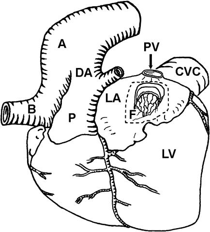
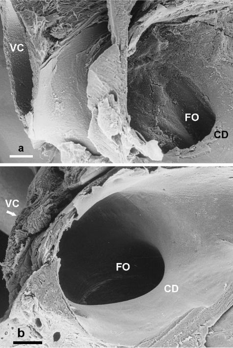
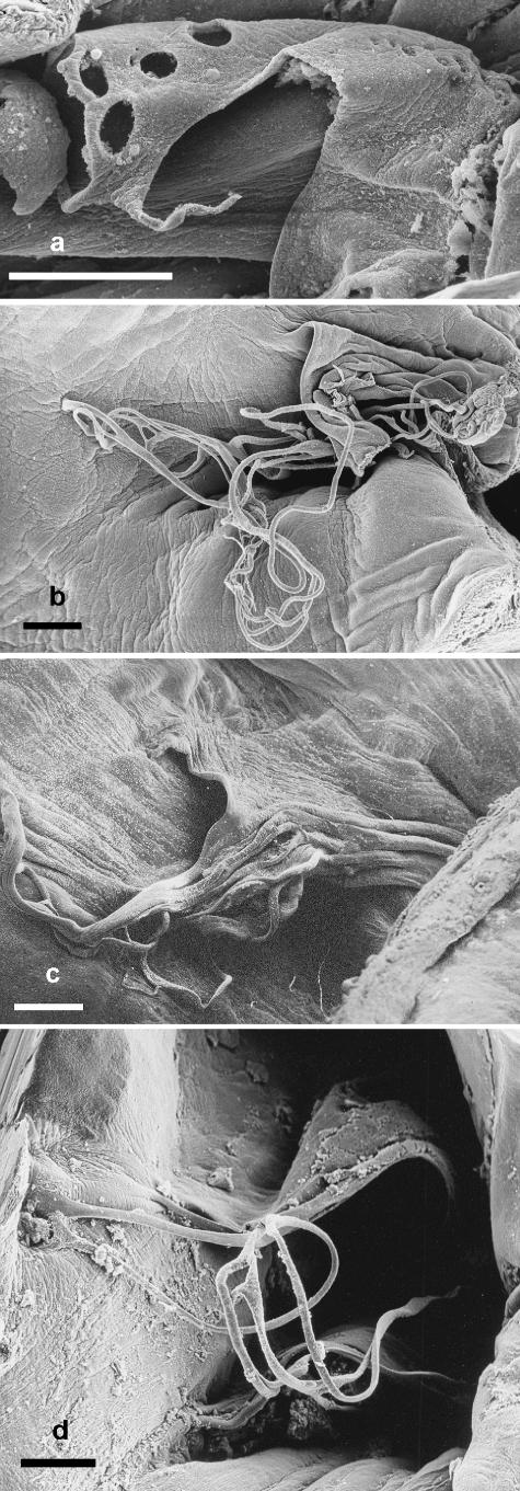
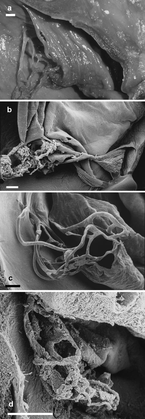
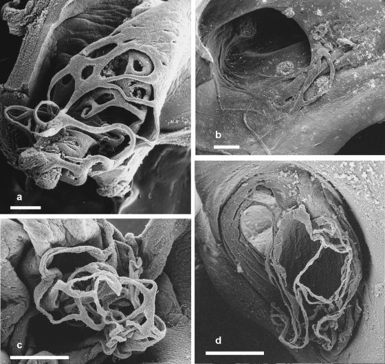
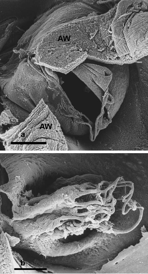
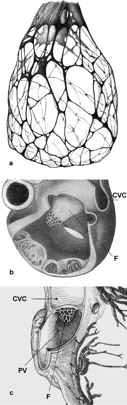
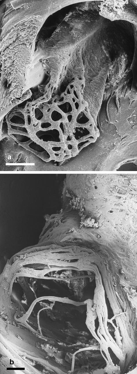
The structure of the cardiac foramen ovale from 17 species representing six cetacean families, the Monodontidae, Phocoenidae, Delphinidae, Ziphiidae, Balaenidae and the Balaenopteridae, was studied using the scanning electron microscope. Eight white whale fetuses (Delphinapterus leucas) and a narwhal fetus (Monodon monoceros) represented the Monodontidae; one fetal and nine neonatal harbour porpoises (Phocoena phocoena) and a finless porpoise fetus (Neophocoena phocoenoides) represented the Phocoenidae; two white-beaked dolphin fetuses (Lagenorhynchus albirostris), four fetal and one neonatal Atlantic white-sided dolphins (Lagenorhynchus acutus), a Risso's dolphin fetus (Grampus griseus), two common bottle-nosed dolphin neonates (Tursiops truncatus), a female short-beaked common dolphin fetus (Delphinus delphis), four killer whale fetuses (Orcinus orca) and two long-finned pilot whale fetuses (Globicephala melas) represented the Delphinidae; two northern bottlenose whale fetuses (Hyperoodon ampullatus) represented the Ziphiidae; one bowhead whale fetus (Balaena mysticetus) represented the Balaenidae and five Common minke whale fetuses (Balaenoptera acutorostrata), one blue whale fetus (Balaenoptera musculus), nine fin whale fetuses (Balaenoptera physalus) and four humpback whale fetuses (Megaptera novaeangliae) represented the Balaenopteridae. The hearts of an additional two incompletely identified toothed and four baleen whale fetuses were also studied. In each species the fold of tissue derived from the cardiac septum primum and subtended by the foramen ovale had the appearance of a short tunnel or sleeve which was fenestrated at its distal end. In the toothed whales the tissue fold was tunnel-shaped with the interatrial septum as the floor whereas in baleen whales it was more sleeve-like. In toothed whales thin threads extended from the fold to insert into the interatrial septum whereas a network of threads covered the distal end of the sleeve in the baleen whales. Similar structures were present in the corresponding cardiac tissues of neonatal Hippopotamidae.
Figures
Similar articles
-
Marine Mammals in the Mediterranean Sea: An Overview.Adv Mar Biol. 2016;75:1-36. doi: 10.1016/bs.amb.2016.08.005. Epub 2016 Sep 28. Adv Mar Biol. 2016. PMID: 27770981
-
Developmental changes of the fore- and hind-limbs in the fetuses of the southern minke whale, Balaenoptera acutorostrata.Anat Anz. 1989;169(2):145-8. Anat Anz. 1989. PMID: 2589636
-
Marine mammal strandings in the New Caledonia region, Southwest Pacific.C R Biol. 2006 Apr;329(4):277-88. doi: 10.1016/j.crvi.2006.01.004. Epub 2006 Feb 17. C R Biol. 2006. PMID: 16644500
-
[Viruses of whales and dolphins].Mikrobiol Z. 1996 Sep-Oct;58(5):100-6. Mikrobiol Z. 1996. PMID: 9044706 Review. Russian.
-
Is it 'boom times' for baleen whales in the Pacific Arctic region?Biol Lett. 2016 Sep;12(9):20160251. doi: 10.1098/rsbl.2016.0251. Biol Lett. 2016. PMID: 27601724 Free PMC article. Review.
Cited by
-
Low incidence of atrial septal defects in nonmammalian vertebrates.Evol Dev. 2020 May;22(3):241-256. doi: 10.1111/ede.12322. Epub 2019 Oct 9. Evol Dev. 2020. PMID: 31597012 Free PMC article.
-
Sequential segmental analysis of the crocodilian heart.J Anat. 2017 Oct;231(4):484-499. doi: 10.1111/joa.12661. Epub 2017 Aug 1. J Anat. 2017. PMID: 28766716 Free PMC article.
-
The Heart of the Killer Whale: Description of a Plastinated Specimen and Review of the Available Literature.Animals (Basel). 2022 Jan 31;12(3):347. doi: 10.3390/ani12030347. Animals (Basel). 2022. PMID: 35158671 Free PMC article.
-
Changes in the Anatomic and Microscopic Structure and the Expression of HIF-1α and VEGF of the Yak Heart with Aging and Hypoxia.PLoS One. 2016 Feb 25;11(2):e0149947. doi: 10.1371/journal.pone.0149947. eCollection 2016. PLoS One. 2016. PMID: 26914488 Free PMC article.
-
Standardization of Dolphin Cardiac Auscultation and Characterization of Heart Murmurs in Managed and Free-Ranging Bottlenose Dolphins (Tursiops truncatus).Front Vet Sci. 2020 Oct 28;7:570055. doi: 10.3389/fvets.2020.570055. eCollection 2020. Front Vet Sci. 2020. PMID: 33240948 Free PMC article.
References
-
- Anon . Out of the Blue: the UK Whale and Dolphin Stranding Scheme. London: Natural History Museum; 2005.
-
- Arnason U, Gullberg A, Janke A. Mitogenomic analyses provide new insights into cetacean origin and evolution. Gene. 2004;333:27–34. - PubMed
-
- Bartholin T. Historiarum anatomicarum rariorum Centuriae II; historia XXV. Amstelodami: 1654.
Publication types
MeSH terms
LinkOut - more resources
Full Text Sources
