

Endothelial cells' activation and apoptosis induced by a subset of antibodies against human cytomegalovirus: relevance to the pathogenesis of atherosclerosis
- PMID: 17534423
- PMCID: PMC1868596
- DOI: 10.1371/journal.pone.0000473
Endothelial cells' activation and apoptosis induced by a subset of antibodies against human cytomegalovirus: relevance to the pathogenesis of atherosclerosis
Abstract
Background: Human cytomegalovirus (hCMV) is involved in the pathogenesis of atherosclerosis. We have previously shown in patients with atherosclerosis that antibodies directed against the hCMV-derived proteins US28 and UL122 are able to induce endothelial cell damage and apoptosis of non-stressed endothelial cells through cross-rection with normally expressed surface molecules. Our aim was to dissect the molecular basis of such interaction and to investigate mechanisms linking innate immunity to atherosclerosis.
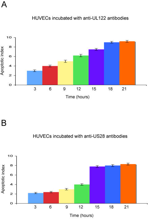
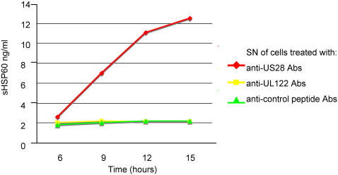
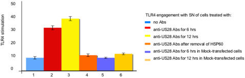
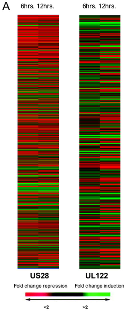
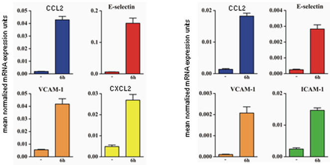
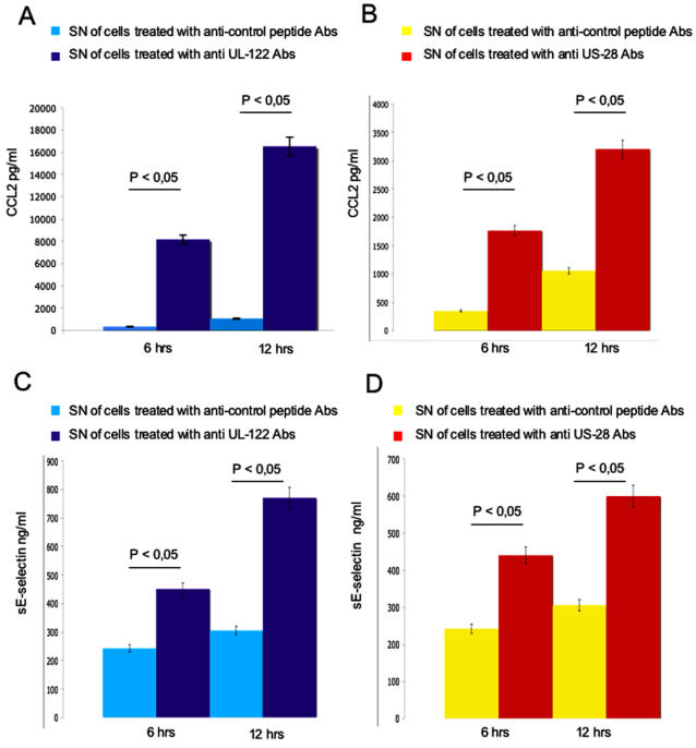
Methodology/principal findings: We analysed the gene expression profiles in endothelial cells stimulated with antibodies affinity-purified against either the UL122 or the US28 peptides using the microarray technology. Microarray results were validated by quantitative PCR and by detection of proteins in the medium. Supernatant of endothelial cells incubated with antibodies was analysed also for the presence of Heat Shock Protein (HSP)60 and was used to assess stimulation of Toll-Like Receptor-4 (TLR4). Antibodies against UL122 and US28 induced the expression of genes encoding for adhesion molecules, chemokines, growth factors and molecules involved in the apoptotis process together with other genes known to be involved in the initiation and progression of the atherosclerotic process. HSP60 was released in the medium of cells incubated with anti-US28 antibodies and was able to engage TLR4.
Conclusions/significance: Antibodies directed against hCMV modulate the expression of genes coding for molecules involved in activation and apoptosis of endothelial cells, processes known to play a pivotal role in the pathogenesis of atherosclerosis. Moreover, endothelial cells exposed to such antibodies express HSP60 on the cell surface and release HSP60 in the medium able to activate TLR4. These data confirm that antibodies directed against hCMV-derived proteins US28 and UL122 purified from patients with coronary artery disease induce endothelial cell damage and support the hypothesis that hCMV infection may play a crucial role in mediating the atherosclerotic process.
Conflict of interest statement
Figures
Similar articles
-
Interaction of antibodies against cytomegalovirus with heat-shock protein 60 in pathogenesis of atherosclerosis.Lancet. 2003 Dec 13;362(9400):1971-7. doi: 10.1016/S0140-6736(03)15016-7. Lancet. 2003. PMID: 14683657
-
Human cytomegalovirus infection and atherothrombosis.J Thromb Thrombolysis. 2012 Feb;33(2):160-72. doi: 10.1007/s11239-011-0662-x. J Thromb Thrombolysis. 2012. PMID: 22161772 Review.
-
Sequencing Directly from Clinical Specimens Reveals Genetic Variations in HCMV-Encoded Chemokine Receptor US28 That May Influence Antibody Levels and Interactions with Human Chemokines.Microbiol Spectr. 2021 Oct 31;9(2):e0002021. doi: 10.1128/Spectrum.00020-21. Epub 2021 Oct 27. Microbiol Spectr. 2021. PMID: 34704798 Free PMC article.
-
Human Cytomegalovirus-Encoded Receptor US28 Is Expressed in Renal Allografts and Facilitates Viral Spreading In Vitro.Transplantation. 2017 Mar;101(3):531-540. doi: 10.1097/TP.0000000000001289. Transplantation. 2017. PMID: 27362315
-
Infections and autoimmunity: role of human cytomegalovirus in autoimmune endothelial cell damage.Lupus. 2015 Apr;24(4-5):419-32. doi: 10.1177/0961203314558677. Lupus. 2015. PMID: 25801885 Review.
Cited by
-
Synergistic interactions between heregulin and peroxisome proliferator-activated receptor-gamma (PPARgamma) agonist in breast cancer cells.J Biol Chem. 2011 Jun 3;286(22):20087-99. doi: 10.1074/jbc.M110.191718. Epub 2011 Apr 5. J Biol Chem. 2011. PMID: 21467033 Free PMC article.
-
Productive Cytomegalovirus Infection Is Associated With Impaired Endothelial Function in ST-Elevation Myocardial Infarction.Am J Med. 2020 Jan;133(1):133-142. doi: 10.1016/j.amjmed.2019.06.021. Epub 2019 Jul 8. Am J Med. 2020. PMID: 31295440 Free PMC article.
-
Viral infection and atherosclerosis.Eur J Clin Microbiol Infect Dis. 2018 Dec;37(12):2225-2233. doi: 10.1007/s10096-018-3370-z. Epub 2018 Sep 5. Eur J Clin Microbiol Infect Dis. 2018. PMID: 30187247 Review.
-
Novel Strategies to Combat CMV-Related Cardiovascular Disease.Pathog Immun. 2020 Sep 20;5(1):240-274. doi: 10.20411/pai.v5i1.382. eCollection 2020. Pathog Immun. 2020. PMID: 33089035 Free PMC article. Review.
-
Zika Virus Transmission Through Blood Tissue Barriers.Front Microbiol. 2019 Jul 4;10:1465. doi: 10.3389/fmicb.2019.01465. eCollection 2019. Front Microbiol. 2019. PMID: 31333605 Free PMC article. Review.
References
-
- Lusis AJ, Mar R, Pajukanta P. Genetics of atherosclerosis. Annu Rev Genomics Hum Genet. 2004;5:189–218. - PubMed
-
- Wilson PW, D'Agostino RB, Levy D, Belanger AM, Silbershatz H, et al. Prediction of coronary heart disease using risk categories. Circulation. 1998;97:837–847. - PubMed
-
- Wich G, Knoflach M, Xu Q. Autoimmune and inflammatory mechanisms in atherosclerosis. Ann Rev Immunol. 2004;22:361–403. - PubMed
-
- Mayr M, Metzler B, Kiechl S, Willeit J, Schett Q, et al. Endothelial cytotoxicity mediated by serum antibodies to heat shock proteins of Escherichia coli and Chlamydia pneumoniae: immune reactions to heat shock proteins as a possible link between infection and atherosclerosis. Circulation. 1999;99:1560–1566. - PubMed
-
- Xiao Q, Mandal K, Schett G, Mayr M, Wick G, et al. Association of serum-soluble heat shock protein 60 with carotid atherosclerosis: clinical significance determined in a follow-up study. Stroke. 2005;12:2571–2576. - PubMed
Publication types
MeSH terms
Substances
LinkOut - more resources
Full Text Sources
Other Literature Sources
Medical
Research Materials
Miscellaneous
