. 2007 Aug;12(6):855-65.
doi: 10.1007/s00775-007-0240-z. Epub 2007 May 30.

Albumin-binding PARACEST agents

Affiliations

Albumin-binding PARACEST agents

M Meser Ali et al. J Biol Inorg Chem. 2007 Aug.

Abstract

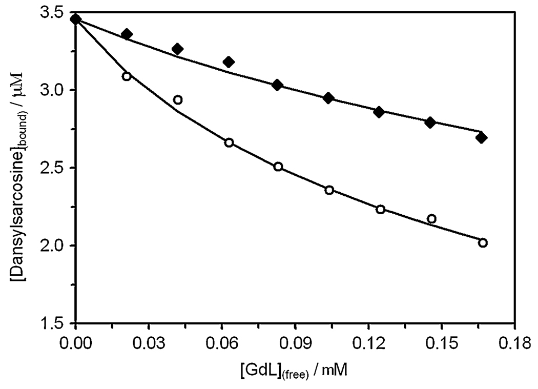
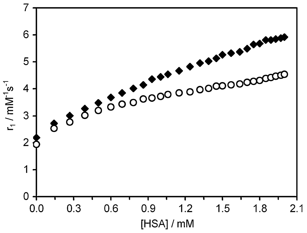
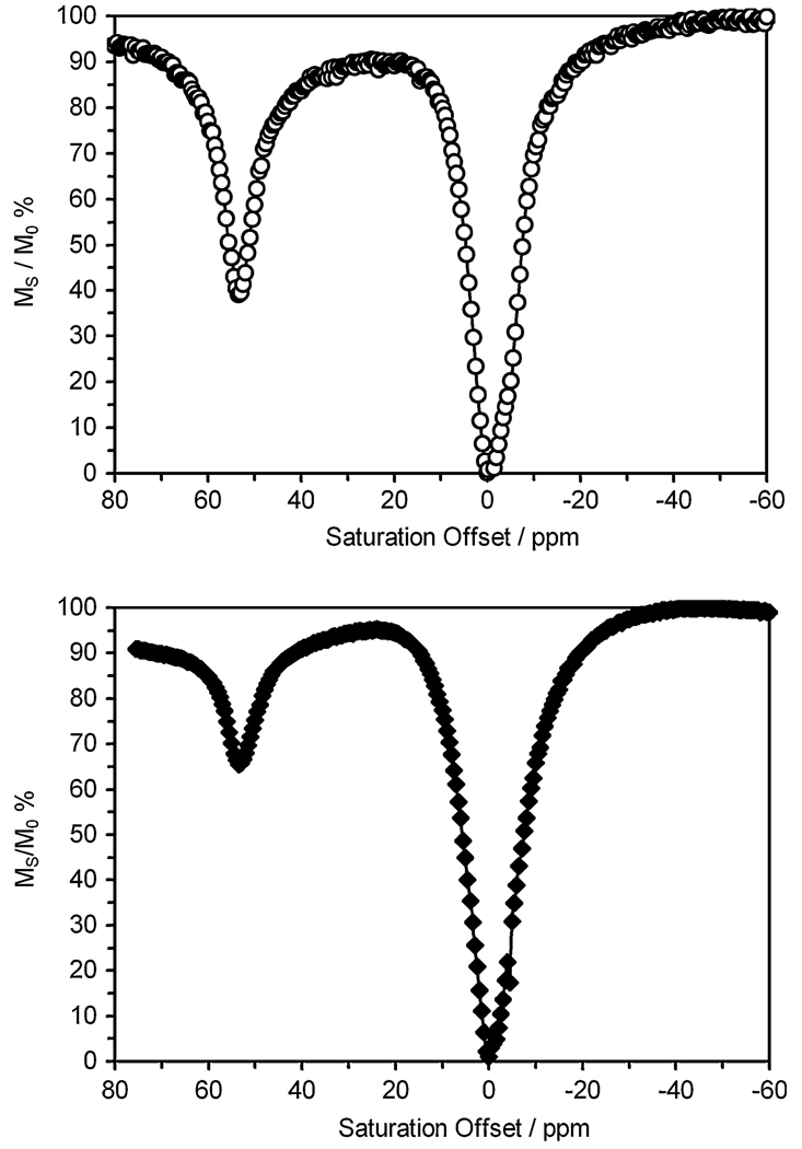
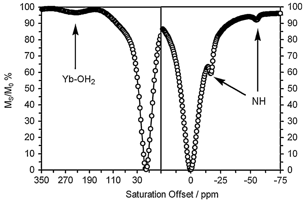
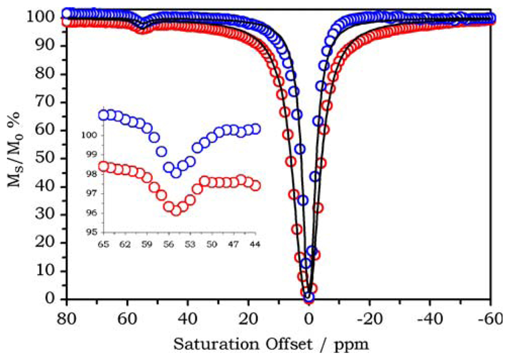
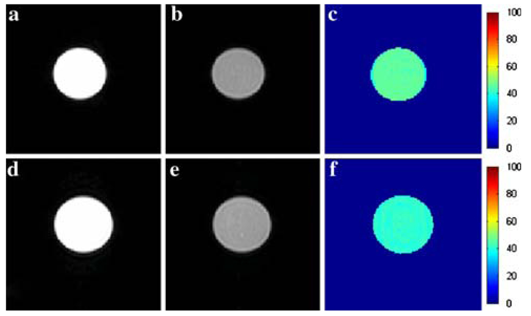
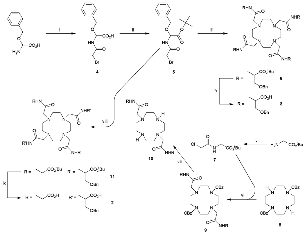
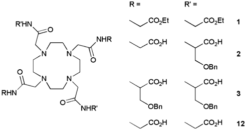
Lanthanide complexes (Eu(3+), Gd(3+) and Yb(3+)) of two different 1,4,7,10-tetraazacyclododecane-1,4,7,10-tetraacetic acid tetraamide derivatives containing two (2) and four (3) O-benzyl-L-serine amide substituents were synthesized and their chemical exchange saturation transfer (CEST) and relaxometric properties were examined in the presence and absence of human serum albumin (HSA). Both Eu2 and Eu3 display a significant CEST effect from a single slowly exchanging Eu(3+)-bound water molecule, making these PARACEST complexes potentially useful as vascular MRI agents. Yb2 also showed a detectable CEST effect from both the Yb(3+)-bound water protons and the exchangeable NH amide protons, making it potentially useful as a vascular pH sensor. Fluorescence displacement studies using reporter molecules indicate that both Gd2 and Gd3 displace dansylsarcosine from site II of HSA with inhibition constants of 32 and 96 microM, respectively, but neither complex significantly displaces warfarin from site I. Water proton relaxation enhancements of 135 and 171% were observed upon binding of Gd2 and Gd3 to HSA, respectively, at 298 K and pH 7.4.


Figures

Fig. 1
Fig. 1
The inhibition of dansylsarcosine binding to human serum albumin (HSA) by Gd2 (circles) and Gd3 (diamonds)
Fig. 2
Fig. 2
Binding of the complexes Gd2 (circles) and Gd3 (diamonds) to HSA has the effect of increasing the longitudinal relaxivity (r1) of each complex. Titrations were performed at 20 MHz, 298 K, in N-(2-hydroxyethyl)piperazine-N′-ethanesulfonic acid buffer, pH 7.4
Fig. 3
Fig. 3
Chemical exchange saturation transfer (CEST) spectra, plotting the solvent water signal intensity (expressed as a percentage of its initial intensity) against presaturation frequency, for a 40 mM aqueous solution of Eu2 (top) and a 20 mM aqueous solution of Eu3 (bottom) at 298 K, irradiation time 2 s, B1 = 26 µT
Fig. 4
Fig. 4
CEST spectra of a 25 mM solution of Yb2 recorded at 500 MHz, pH 7.4, and 298 K. On the left is shown the downfield region of the spectrum recorded with an irradiation time of 4 s, B1 = 6.5 µT; a small CEST peak arising from the coordinated water molecule is clearly visible. On the right is shown the upfield region for an irradiation time of 1 s, B1 = 26 µT; CEST peaks arising from the amide NH protons can be seen
Fig. 5
Fig. 5
The CEST spectra of 0.75 mM Eu2 in phosphate-buffered saline (PBS) recorded in the absence (blue) and presence (red) of 0.75 mM HSA. B0 = 400 MHz, B1 = 19 µT, irradiation time 6 s, 298 K
Fig. 6
Fig. 6
Images of 20 mM solutions of Eu2 in the absence and presence of 5% HSA. Left (a, d), center (b, e), and right (c, f) columns represent the off-resonance (−54 ppm), on-resonance (54 ppm), and the percentage CEST enhancement maps, respectively. Samples in the top row contained 20 mM Eu2 in PBS, while samples in the bottom row contained in addition 5% HSA (Buminate®). Each image was collected using a standard spin-echo sequence with a 6-s presaturation pulse applied at the indicated offset
Scheme 1
Scheme 1
The synthetic route to ligands 2 and 3. Reagents and conditions: i BrCH2COBr/K2CO3/NaOH/273 K; ii tert-butyl trichloroacetimidate/BF3·OEt/tetrahydrofuran/273 K; iii cyclen/iPr2NEt/MeCN/328 K; iv trifluoroacetic acid (TFA); v ClCH2COCl/K2CO3/H2O/CH2Cl2/273 K; vi K2CO3/MeCN/333 K; vii H2/Pd on C; viii iPr2NEt/MeCN/333 K; ix TFA
Structure 1
Structure 1

References

    1. Merbach AE, Toth E. The chemistry of contrast agents in medical magnetic resonance imaging. Chichester: Wiley; 2001.
    1. Caravan P, Ellison JJ, McMurry TJ, Lauffer RB. Chem Rev. 1999;99:2293–2352. - PubMed
    1. Ward KM, Balaban RS. Magn Reson Med. 2000;44:799–802. - PubMed
    1. Terreno E, Castelli Daniela D, Cravotto G, Milone L, Aime S. Invest Radiol. 2004;39:235–243. - PubMed
    1. Goffeney N, Bulte JWM, Duyn J, Bryant LH, Jr, van Zijl PCM. J Am Chem Soc. 2001;123:8628–8629. - PubMed

Publication types

MeSH terms