. 2007 Jul;5(7):e163.
doi: 10.1371/journal.pbio.0050163. Epub 2007 May 29.

All beta cells contribute equally to islet growth and maintenance

Affiliations

All beta cells contribute equally to islet growth and maintenance

Kristen Brennand et al. PLoS Biol. 2007 Jul.

Abstract

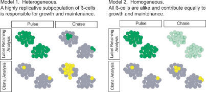
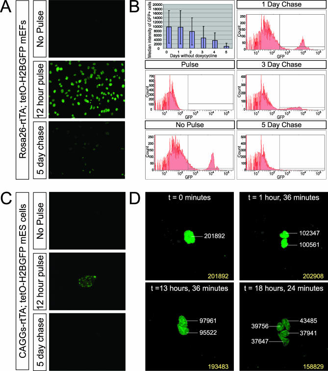
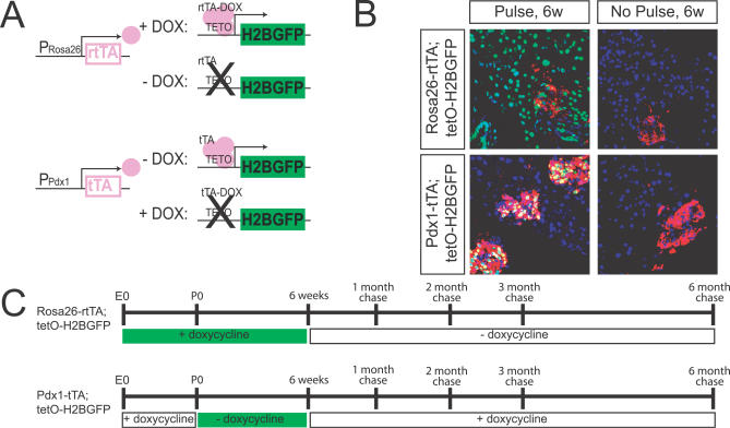
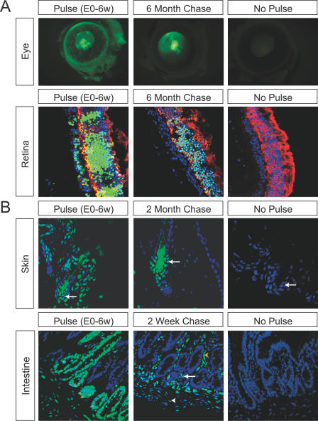
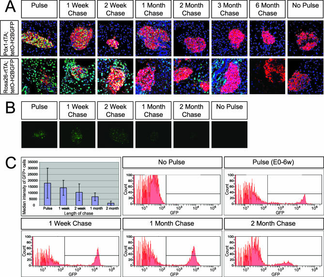
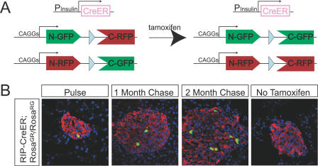
In healthy adult mice, the beta cell population is not maintained by stem cells but instead by the replication of differentiated beta cells. It is not known, however, whether all beta cells contribute equally to growth and maintenance, as it may be that some cells replicate while others do not. Understanding precisely which cells are responsible for beta cell replication will inform attempts to expand beta cells in vitro, a potential source for cell replacement therapy to treat diabetes. Two experiments were performed to address this issue. First, the level of fluorescence generated by a pulse of histone 2B-green fluorescent protein (H2BGFP) expression was followed over time to determine how this marker is diluted with cell division; a uniform loss of label across the entire beta cell population was observed. Second, clonal analysis of dividing beta cells was completed; all clones were of comparable size. These results support the conclusion that the beta cell pool is homogeneous with respect to replicative capacity and suggest that all beta cells are candidates for in vitro expansion. Given similar observations in the hepatocyte population, we speculate that for tissues lacking an adult stem cell, they are replenished equally by replication of all differentiated cells.


Conflict of interest statement

Competing interests. The authors have declared that no competing interests exist.

Figures

Figure 1
Figure 1. Two Possible Models for the Growth and Maintenance of Pancreatic β Cells Are Predicted, Given the Lack of an External Stem Cell Pool
Two approaches were used to study replication of β cells. Pulse–chase analysis follows the loss of the H2BGFP label (green) with β cell division, while clonal analysis follows the generation of (yellow) clones from individual β cells. Model 1: the β cell population is heterogeneous, composed of fast- and slow-dividing subpopulations. This model predicts that highly replicative β cells will lose the H2BGFP label quickly and generate large clones, while slowly dividing β cells will fail to dilute the H2BGFP label and generate small clones. Model 2: the β cell population is homogeneous, and all β cells divide at the same rate. This model predicts that all β cells will lose the H2BGFP label uniformly, and that all clones identified through clonal analysis will be of comparable size.
Figure 2
Figure 2. In Vitro Characterization of Dilution of Tetracycline-Inducible H2BGFP with Cell Division
(A) Fluorescent images of Rosa26-rtTA; tetO-H2BGFP mEFs pulsed with doxycycline and cultured in the absence of doxycycline. Original magnification, 100×. (B) FACS plots of Rosa26-rtTA; tetO-H2BGFP mEFs pulsed with doxycycline and cultured in the absence of doxycycline. Median intensity of GFP-positive cells versus days cultured without doxycycline shown in graph. (C) Fluorescent images of CAGGs-rtTA; tetO-H2BGFP mES cells pulsed with doxycycline and cultured in the absence of doxycycline. Original magnification, 100×. (D) Time-lapse confocal images of a single CAGGs-rtTA; tetO-H2BGFP mES cell undergoing two rounds of cell division. Integrated pixel intensity shown in white text; total integrated pixel intensity of all cells shown in yellow text. Original magnification, 400×.
Figure 3
Figure 3. Experimental Schematic for Use of Tetracycline-Inducible H2BGFP To Identify LRCs In Vivo
(A) Schematic for Rosa26-rtTA; tetO-H2BGFP and Pdx1-tTA; tetO-H2BGFP expression. (B) Doxycycline-dependent expression of Rosa26-rtTA; tetO-H2BGFP and Pdx1-tTA; tetO-H2BGFP expression in the pancreas. Insulin expression is shown in red. Up to 80% of ß cells are labeled following the pulse period; no ß cells are labeled in the absence of pulse. Original magnification, 400×. (C) Timeline for Rosa26-rtTA; tetO-H2BGFP and Pdx1-tTA; tetO-H2BGFP pulse–chase experiments.
Figure 4
Figure 4. Identification of LRCs in Postmitotic Cells and Stem Cell Populations Indicates Stability of H2BGFP Fluorescence
(A) LRCs detected in the postmitotic photoreceptor cells of the retina after expression of Rosa26-rtTA; tetO-H2BGFP from E0–E6 wk. Top row: H2BGFP fluorescence in the eye persists at least 6 mo following initial pulse. Original magnification, 20×. Bottom row: label retention in the retina is restricted to postmitotic photoreceptor cells, labeled in red with anti-recoverin. Original magnification, 400×. All images are exposure matched. (B) LRCs detected in the adult skin and intestine after expression of Rosa26-rtTA; tetO-H2BGFP from E0–E6 wk. In the skin, bulge stem cells are labeled with an arrow. In the intestine, a putative crypt cell is marked with an arrow. White arrowheads mark smooth muscle, and yellow arrowheads label enteric neurons. Images are exposure matched. Original magnification: skin, 630×; intestine, 400×.
Figure 5
Figure 5. Uniform Loss of Label in Adult β Cells Following Pulse–Chase with Pdx1-tTA; tetO-H2BGFP and Rosa26-rtTA; tetO-H2BGFP Mice
(A) Label retention present in the adult pancreas (insulin expression shown in red) following a chase period of up to 6 mo. Exposure-matched images. Original magnification, 400×. (B) Whole islets of Rosa26-rtTA; tetO-H2BGFP pulse–chase animals prior to dissociation and FACS. Exposure-matched images. Original magnification, 100×. (C) FACS plots of dissociated islets of Rosa26-rtTA; tetO-H2BGFP pulse–chase animals. Median intensity of GFP-positive cells versus length of chase shown in graph.
Figure 6
Figure 6. Clonal Analysis of β cells
(A) Schematic for the RIP-CreER; Rosa26GR/Rosa26RG pulse–chase experiments. (B) RIP-CreER; Rosa26GR/Rosa26RG mice were pulsed with a tamoxifen injection between 4–8 wk of age. Shown here are sections of typical β cell clones (green) within 4 d of the pulse and after 1 and 2 mo of chase. β cells are identified by insulin staining (red). Original magnification, 400×.

Comment in

References

    1. Cheng H, Leblond CP. Origin, differentiation and renewal of the four main epithelial cell types in the mouse small intestine. V. Unitarian theory of the origin of the four epithelial cell types. Am J Anat. 1974;141:537–561. - PubMed
    1. Oshima H, Rochat A, Kedzia C, Kobayashi K, Barrandon Y. Morphogenesis and renewal of hair follicles from adult multipotent stem cells. Cell. 2001;104:233–245. - PubMed
    1. Spangrude GJ, Heimfeld S, Weissman IL. Purification and characterization of mouse hematopoietic stem cells. Science. 1988;241:58–62. - PubMed
    1. Reynolds BA, Weiss S. Generation of neurons and astrocytes from isolated cells of the adult mammalian central nervous system. Science. 1992;255:1707–1710. - PubMed
    1. Doetsch F, Caille I, Lim DA, Garcia-Verdugo JM, Alvarez-Buylla A. Subventricular zone astrocytes are neural stem cells in the adult mammalian brain. Cell. 1999;97:703–716. - PubMed

Publication types

MeSH terms