. 2007 May 29:7:49.
doi: 10.1186/1471-2180-7-49.

Inhibition of apoptosis prevents West Nile virus induced cell death

Affiliations

Inhibition of apoptosis prevents West Nile virus induced cell death

Malte C Kleinschmidt et al. BMC Microbiol. .

Abstract

Background: West Nile virus (WNV) infection can cause severe meningitis and encephalitis in humans. Apoptosis was recently shown to contribute to the pathogenesis of WNV encephalitis. Here, we used WNV-infected glioma cells to study WNV-replication and WNV-induced apoptosis in human brain-derived cells.

Results: T98G cells are highly permissive for lytic WNV-infection as demonstrated by the production of infectious virus titre and the development of a characteristic cytopathic effect. WNV replication decreased cell viability and induced apoptosis as indicated by the activation of the effector caspase-3, the initiator caspases-8 and -9, poly(ADP-ribose)polymerase (PARP) cleavage and the release of cytochrome c from the mitochondria. Truncation of BID indicated cross-talk between the extrinsic and intrinsic apoptotic pathways. Inhibition of the caspases-8 or -9 inhibited PARP cleavage, demonstrating that both caspases are involved in WNV-induced apoptosis. Pan-caspase inhibition prevented WNV-induced apoptosis without affecting virus replication.

Conclusion: We found that WNV infection induces cell death in the brain-derived tumour cell line T98G by apoptosis under involvement of constituents of the extrinsic as well as the intrinsic apoptotic pathways. Our results illuminate the molecular mechanism of WNV-induced neural cell death.


Figures
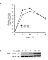
Figure 1
Figure 1
T98G cells are permissive for WNV infection. Production of infectious virus titres (A) and accumulation of viral envelope antigen (B) in T98G cell cultures infected with WNV at different time points p.i. Values represent the mean (± SD) of three different experiments. The blot shown is representative for a set of three different experiments.
Figure 2
Figure 2
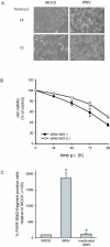
Effects of WNV on T98G cell viability. WNV-induced CPE (MOI 1) (A), cell viability of WNV-infected T98G cell cultures relative to mock-infected T98G cells (B) and virus induced PARP cleavage (MOI 1) (C) in infected cells. Values represent the mean (± SD) of three different experiments and pictures shown are representative for a set of three different experiments. *p < 0.05 compared to mock-infected cultures.
Figure 3
Figure 3
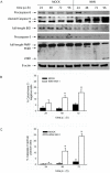
WNV infection induces caspase activation and cytochrome c release. Cellular levels of procaspase-8, cleaved caspase-9, BID and procaspase-3 during WNV-infection (A), release of cytochrome c from mitochondria into the cytoplasma (B) and caspase-3 activation (C) in WNV infected cells (all MOI 1). The band in A marked with * resulted from unspecific antibody binding. Blots shown are representative for a set of three different experiments. Values represent mean (± SD) from three independent experiments. *p < 0.05 compared to mock-infected cultures.
Figure 4
Figure 4
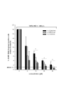
Caspase inhibitor peptides inhibit WNV-induced PARP-cleavage. Effect on WNV-induced PARP cleavage of peptides z-IETD-fmk (caspase-8 inhibitor), z-LEHD-fmk (caspase-9 inhibitor) and z-VAD-fmk (pancaspase inhibitor) at 48 h p.i. Results are expressed as percentage of values obtained from WNV-infected cultures (MOI 1) without any inhibitor added. Values represent mean (± SD) from three independent experiments. *p < 0.05 compared to untreated virus control.
Figure 5
Figure 5
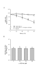
Prevention of WNV-induced cell death by pan-caspase inhibition. Inhibition of virus-induced cell death indicated by trypan blue staining of WNV (MOI 0.1)-infected T98G cell cultures by pan-caspase inhibitor z-VAD-fmk (100 μM) relative to mock-infected cells (A) and the effect of different z-VAD-fmk concentrations on infectious virus titre production (TCID50/ml, 48 h p.i.) after infection at MOI 0.1 (B). Values represent mean (± SD) from three independent experiments. *p < 0.05 compared to untreated virus control.

Similar articles

Cited by

References

    1. Campbell GL, Marfin AA, Lanciotti RS, Gubler DJ. West Nile virus. Lancet Infect Dis. 2002;2:519–529. doi: 10.1016/S1473-3099(02)00368-7. - DOI - PubMed
    1. Granwehr BP, Lillibridge KM, Higgs S, Mason PW, Aronson JF, Campbell GA, Barrett AD. West Nile virus: where are we now? Lancet Infect Dis. 2004;4:547–556. doi: 10.1016/S1473-3099(04)01128-4. - DOI - PubMed
    1. Nash D, Mostashari F, Fine A, Miller J, O'Leary D, Murray K, Huang A, Rosenberg A, Greenberg A, Sherman M, Wong S, Layton M, West Nile Outbreak Response Working Group The outbreak of West Nile virus infection in the New York City area in 1999. N Engl J Med. 2001;344:1807–1814. doi: 10.1056/NEJM200106143442401. - DOI - PubMed
    1. Fan TJ, Han LH, Cong RS, Liang J. Caspase family proteases and apoptosis. Acta Biochim Biophys Sin (Shanghai) 2005;37:719–727. doi: 10.1111/j.1745-7270.2005.00108.x. - DOI - PubMed
    1. Chu JJ, Ng ML. The mechanism of cell death during West Nile virus infection is dependent on initial infectious dose. J Gen Virol. 2003;84:3305–3314. doi: 10.1099/vir.0.19447-0. - DOI - PubMed

Publication types