. 2007 May 30:8:45.
doi: 10.1186/1471-2199-8-45.

Regulation of p73 by Hck through kinase-dependent and independent mechanisms

Affiliations

Regulation of p73 by Hck through kinase-dependent and independent mechanisms

Preeti Paliwal et al. BMC Mol Biol. .

Abstract

Background: p73, a p53 family member is a transcription factor that plays a role in cell cycle, differentiation and apoptosis. p73 is regulated through post translational modifications and protein interactions. c-Abl is the only known tyrosine kinase that phosphorylates and activates p73. Here we have analyzed the role of Src family kinases, which are involved in diverse signaling pathways, in regulating p73.

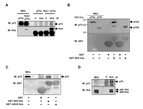
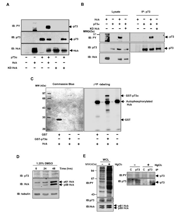
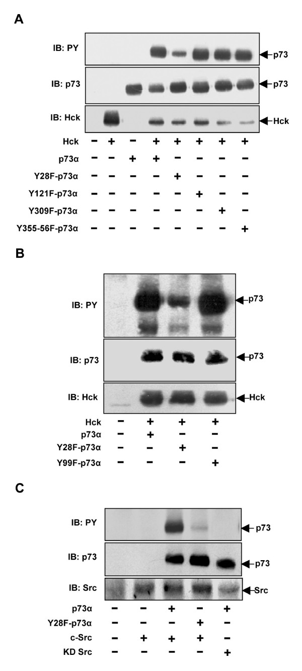
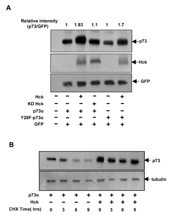
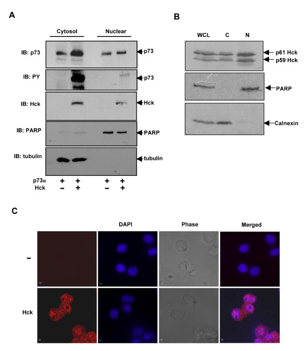
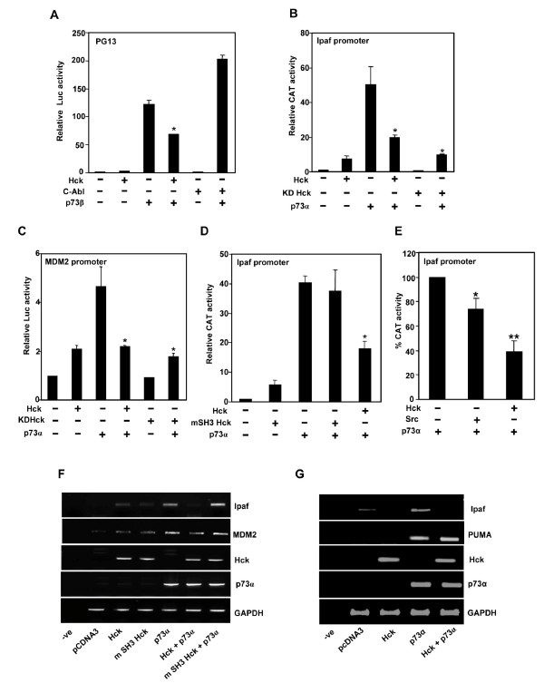
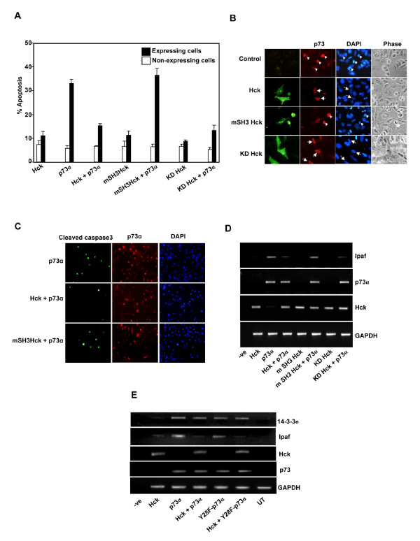
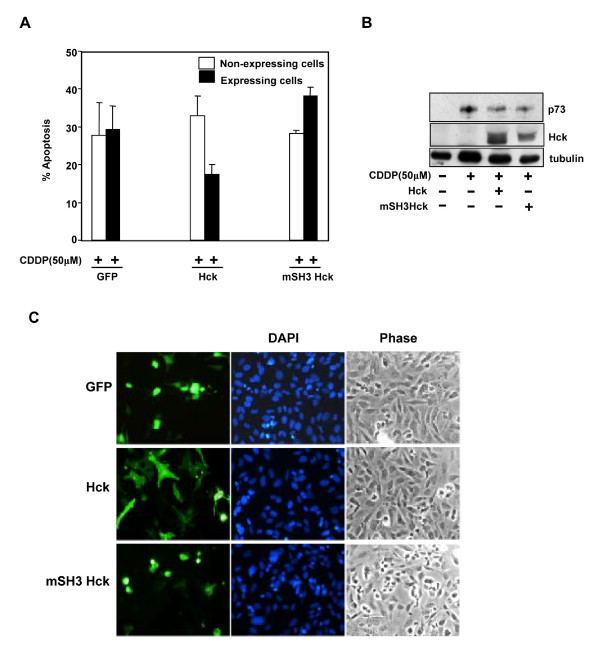
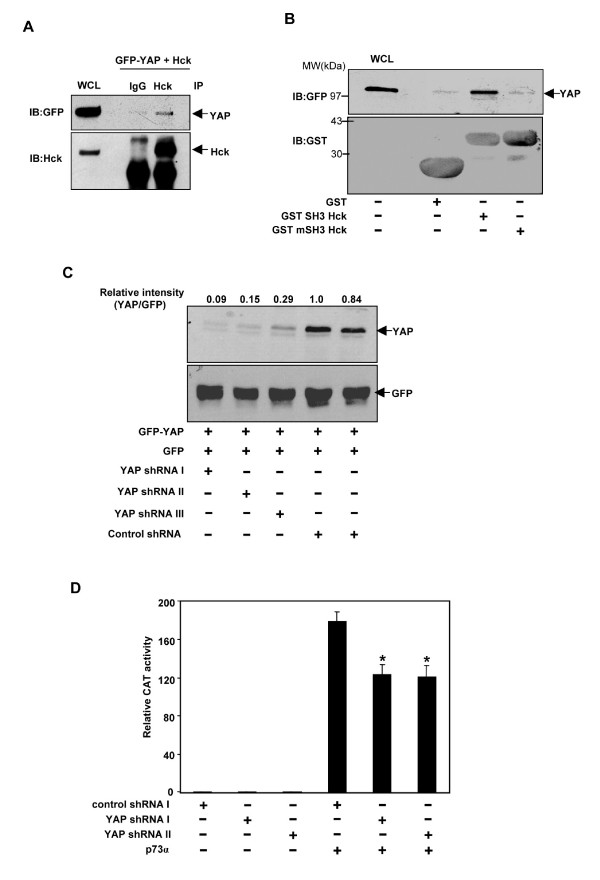
Results: Exogenously expressed as well as cellular Hck and p73 interact in vivo. In vitro binding assays show that SH3 domain of Hck interacts with p73. Co-expression of p73 with Hck or c-Src in mammalian cells resulted in tyrosine phosphorylation of p73. Using site directed mutational analysis, we determined that Tyr-28 was the major site of phosphorylation by Hck and c-Src, unlike c-Abl which phosphorylates Tyr-99. In a kinase dependent manner, Hck co-expression resulted in stabilization of p73 protein in the cytoplasm. Activation of Hck in HL-60 cells resulted in tyrosine phosphorylation of endogenous p73. Both exogenous and endogenous Hck localize to the nuclear as well as cytoplasmic compartment, just as does p73. Ectopically expressed Hck repressed the transcriptional activity of p73 as determined by promoter assays and semi-quantitative RT-PCR analysis of the p73 target, Ipaf and MDM2. SH3 domain- dependent function of Hck was required for its effect on p73 activity, which was also reflected in its ability to inhibit p73-mediated apoptosis. We also show that Hck interacts with Yes associated protein (YAP), a transcriptional co-activator of p73, and shRNA mediated knockdown of YAP protein reduces p73 induced Ipaf promoter activation.

Conclusion: We have identified p73 as a novel substrate and interacting partner of Hck and show that it regulates p73 through mechanisms that are dependent on either catalytic activity or protein interaction domains. Hck-SH3 domain-mediated interactions play an important role in the inhibition of p73-dependent transcriptional activation of a target gene, Ipaf, as well as apoptosis.


Figures

Figure 1
Figure 1
Hck interacts with p73α in vitro and in vivo. (A) Cos1 cells transiently expressing HA-p73α alone or with Hck were immunoprecipitated with control rabbit IgG (C) or Hck polyclonal antibody. The immunoprecipitates were resolved by SDS-PAGE and subjected to western blotting to detect p73 and Hck. (B) GST fusion proteins bound to Glutathione-Sepharose beads were incubated with lysates of Cos1 cells overexpressing p73α and p73δ. Bound proteins were subjected to western blotting to detect p73. (C) GST-SH3 Hck and GST-mSH3 Hck recombinant fusion proteins bound to Glutathione-Sepharose beads were incubated with lysates of Cos1 cells overexpressing p73α and bound proteins were subjected to western blotting with p73 antibody. (D) Lysates of differentiated HL-60 cells (DMSO 1.25%) were immunoprecipitated with control rabbit IgG or Hck antibodies and subjected to western blotting for p73 and Hck. (WCL indicates whole cell lysate).
Figure 2
Figure 2
Hck phosphorylates p73α in vivo and in vitro. (A) Whole cell lysates prepared from Cos1 cells transfected with HA-p73α (1.6 μg) with and without Hck (0.4 μg) expression constructs in the ratio of 4:1 were subjected to immunoblotting with pTyr, p73 and Hck antibodies. (B) Cos1 cells transfected with indicated expression constructs were subjected to immunoprecipitation with p73 antibody and immunoprecipitates analyzed for pTyr, p73 and Hck by western blotting. The amount of DNA was kept constant by addition of control vector pcDNA3. (C) Purified Hck protein (80 nM) was incubated with GST, GST-p73α and alone with γ32P-ATP for 30 minutes at 37°C in an in vitro kinase assay. The proteins were analyzed by SDS-PAGE and stained with commassie blue (left panel). The gel was then dried and phosphorylated proteins visualized by phosphor imaging (right panel). (D) Western blot showing the endogenous protein levels of p73 and Hck upon differentiation with DMSO in HL-60 cells. Tubulin expression was determined as a loading control. (E) Endogenous p73 gets phosphorylated on tyrosine upon activation of Hck. Western blot showing the phosphotyrosine content of cellular proteins and levels of endogenous p73 and Hck upon HgCl2 treatment in differentiated HL-60 cells (left panel). Lysates of diferentiated HL-60 cells treated with or without HgCl2 were subjected to immunoprecipitation with control (rabbit IgG) or p73 (rabbit polyclonal) antibody and western blotting performed with anti-phosphotyrosine antibody (right panel, upper portion) Immunoprecipitated p73 is shown in right panel, lower portion.
Figure 3
Figure 3
Tyr-28 is the major site of phosphorylation on p73 upon Hck co-expression. (A, B) Cos1 cells transfected with indicated combinations of p73 and Hck plasmids were analyzed by western blotting for pTyr. The same blot was reprobed for p73 and Hck. (C) Src tyrosine kinase phosphorylates p73α on Tyr-28 residue. Cos1 cells transfected with p73α, Y28F-p73α or c-Src and KD-Src were subjected to western blotting for pTyr, p73 and Src.
Figure 4
Figure 4
Hck stabilizes p73α dependent on its kinase activity. (A) Hela cells transfected with GFP (50 ng) and Hck or KDHck(200 ng) in the presence of p73α or Y28F-p73α (50 ng) were subjected to western blotting for p73, Hck and GFP. GFP was used as a transfection efficiency control. (B) Hela cells transfected with p73α or p73α and Hck together were treated with cycloheximide (50 μg/ml), harvested at the indicated time periods and immunoblotted for p73 and tubulin. The amount of DNA in transfections was kept constant by the addition of control vector pcDNA3.
Figure 5
Figure 5
Sub-cellular localization of Hck and p73α. (A) Cos1 cells transiently transfected with p73α or Hck and p73α were fractionated into nuclear and cytosolic fractions and analysed by immunoblotting for pTyr, p73 and Hck. The purity of nuclear and cytosolic fractions was confirmed by immunoblotting for PARP and α-tubulin. (B) Endogenous Hck in HL-60 cells localizes to both cytosolic and nuclear fractions. HL-60 cells were fractionated into nuclear and cytosolic (post nuclear fraction) fractions and immunoblotted using Hck antibody. The purity of fractions was confirmed by immunoblotting with PARP and Calnexin. WCL indicates whole cell lysates. (C) Localization of endogenous Hck in the nucleus by immunostaining. HL-60 cells were immunostained with Hck antibody after 24 hours of differentiation by TPA (10 ng/ml) and analysed by confocal microscopy. The image shown is the central section passing through nucleus. The cells were also stained without primary antibody, which served as a control.
Figure 6
Figure 6
Hck inhibits the transcriptional activity of p73α and β isoforms, dependent on its SH3 domain. (A-D) Hela cells transfected with pCMV-βgal (50 ng) and different promoter constructs (100 ng) along with p73α or p73β and Hck or KD-Hck were subjected to either luciferase or CAT activity measurements. The amount of DNA was kept constant in all transfections to 400 ng by adding pcDNA3. Hck, KD-Hck, mSH3-Hck and c-Abl plasmids were used at 100 ng for different promoter constructs. The amount of p73α used was 2 ng for Ipaf-CAT promoter and 50 ng for MDM2-Luc promoter. The amount of p73β used was 10 ng for PG13-Luc promoter. Relative reporter activities were calculated after normalizing with β-galactosidase activities. Data presented here are mean ± S.D. of at least three independent experiments, *p < 0.01.(E) Effect of c-Src on p73 induced Ipaf promoter transctivation. HeLa cells transfected with Ipaf promoter construct along with p73α (2 ng) and c-Src or Hck (100 ng) were subjected to CAT activity measurements. Data presented here are mean ± S.D. of at least three independent experiments, *p < 0.05, **p < 0.01. (F, G) Total RNA isolated from Hela cells transfected with HA-p73α (200 ng) or Hck (1300 ng) and mSH3 Hck (1300 ng) were subjected to RT-PCR analysis for Ipaf, MDM2 and PUMA gene expression. p73α and Hck mRNA levels were determined for transfection efficiency control whereas GAPDH mRNA levels were used as an internal control.
Figure 7
Figure 7
Hck inhibits p73α-induced apoptosis. (A). SAOS-2 cells transfected with p73α and Hck, mSH3-Hck or KD-Hck in the ratio of 1:3 were immunostained for p73 and Hck and the percentage of apoptotic cells was scored among expressing and non-expressing cells using morphological criteria. Data represent mean ± S.D. of at least three independent experiments performed on duplicate coverslips. (B) Panels show the morphological features of p73 and Hck expressing cells. Arrowhead shows apoptotic cells and arrows indicate healthy cells. (C) SAOS-2 cells transfected with p73α and Hck or mSH3-Hck in the ratio of 1:3 were immunostained for p73 and cleaved caspase-3 expression. (D) Total RNA isolated from SAOS-2 cells transfected with HA-p73α (200 ng) with or without Hck (1300 ng), mSH3 Hck (1300 ng) and KD Hck (1300 ng) were subjected to RT-PCR analysis as described in Fig. 7E. (E) p73α transactivation function is independent of Tyr-28 phosphorylation. RT-PCR analysis was carried out using RNA isolated from SAOS-2 cells after transfection with indicated expression constructs. Expression levels of gene products were determined using appropriate primers. (UT indicates untransfected).
Figure 8
Figure 8
Hck inhibits cisplatin induced apoptosis. (A) SAOS2 cells transfected with GFP, Hck or mSH3 Hck (400 ng) were treated with 50 μM cisplatin (CDDP) for 24 hours and immunostained for Hck. The percentage of apoptotic cells was scored among expressing and non-expressing cells using morphological criteria. Data represent mean ± S.D. of at least three independent experiments. (B) SAOS2 cells transfected with Hck or mSH3 Hck (400 ng) were treated with 50 μM cisplatin (CDDP) for 24 hours and whole cell lysates were subjected to western blotting with p73 (mouse monoclonal, Imgenex) and Hck antibody. Tubulin was used as loading control. (C) Panels show the expression of GFP, Hck and mSH3 Hck expressing cells.
Figure 9
Figure 9
Hck interacts with YAP and requirement of YAP for transactivation of Ipaf by p73. (A) Cos1 cells transiently expressing GFP-YAP with Hck were immunoprecipitated with control rabbit IgG (C) or Hck polyclonal antibody. The immunoprecipitates were resolved by SDS-PAGE and subjected to western blotting to detect YAP and Hck. (B) Extract of Cos1 cells transfected with GFP-YAP was incubated with GST, GST-SH3Hck, GST-mSH3-Hck recombinant proteins bound to Glutathione Sepharose beads. Proteins bound to these beads were immunoblotted with GFP and GST antibodies. (C) YAP-shRNA downregulates GFP-YAP expression. Hela cells transfected with GFP-YAP (50 ng) with either control shRNA I and II (200 ng) or YAPshRNA I, II and III (200 ng) subjected to immunoblotting with anti-GFP antibody. GFP (50 ng) was used as transfection efficiency control. (D). Effect of YAP shRNA on p73α mediated Ipaf promoter transactivation. HeLa cells transiently transfected with YAPshRNA I, II (200 ng) or control shRNA I (200 ng) in the presence or absence of p73α (2 ng) along with Ipaf-CAT promoter construct (150 ng) and pCMV-β Gal (50 ng) were subjected to CAT activity measurements.

Similar articles

Cited by

References

    1. Kaghad M, Bonnet H, Yang A, Creancier L, Biscan JC, Valent A, Minty A, Chalon P, Lelias JM, Dumont X, et al. Monoallelically expressed gene related to p53 at 1p36, a region frequently deleted in neuroblastoma and other human cancers. Cell. 1997;90:809–819. doi: 10.1016/S0092-8674(00)80540-1. - DOI - PubMed
    1. Ozaki T, Nakagawara A. p73, a sophisticated p53 family member in the cancer world. Cancer Sci. 2005;96:729–737. doi: 10.1111/j.1349-7006.2005.00116.x. - DOI - PMC - PubMed
    1. Melino G, De LV, Vousden KH. p73: Friend or foe in tumorigenesis. Nat Rev Cancer. 2002;2:605–615. doi: 10.1038/nrc861. - DOI - PubMed
    1. Harms K, Nozell S, Chen X. The common and distinct target genes of the p53 family transcription factors. Cell Mol Life Sci. 2004;61:822–842. doi: 10.1007/s00018-003-3304-4. - DOI - PMC - PubMed
    1. Jost CA, Marin MC, Kaelin WG., Jr p73 is a simian [correction of human] p53-related protein that can induce apoptosis. Nature. 1997;389:191–194. doi: 10.1038/38298. - DOI - PubMed

Publication types

MeSH terms