. 2007 Jun 12;104(24):10252-7.
doi: 10.1073/pnas.0703676104. Epub 2007 May 29.

Stepwise proteolysis liberates tau fragments that nucleate the Alzheimer-like aggregation of full-length tau in a neuronal cell model

Affiliations

Stepwise proteolysis liberates tau fragments that nucleate the Alzheimer-like aggregation of full-length tau in a neuronal cell model

Y P Wang et al. Proc Natl Acad Sci U S A. .

Abstract

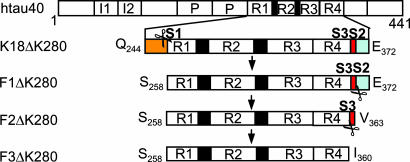
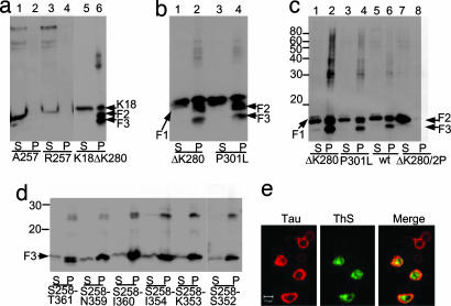
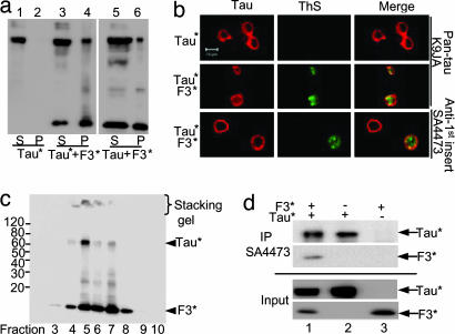
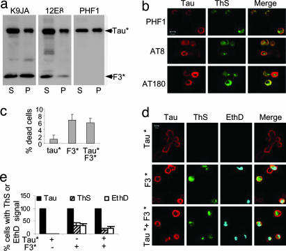
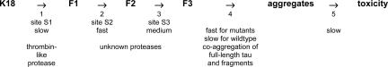
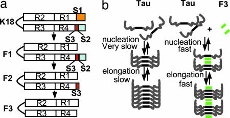
Tau is a highly soluble protein, yet it aggregates abnormally in Alzheimer's disease. Here, we address the question of proteolytic processing of tau and the nucleation of aggregates by tau fragments. We show in neuronal cell models that fragments of the repeat domain of tau containing mutations of FTDP17 (frontotemporal dementia with parkinsonism linked to chromosome 17), produced by endogenous proteases, can induce the aggregation of full-length tau. Fragments are generated by successive cleavages, first N-terminally between K257 and S258, then C-terminally around residues 353-364; conversely, when the N-terminal cleavage is inhibited, no fragmentation and aggregation takes place. The C-terminal truncation and the coaggregation of fragments with full-length tau depends on the propensity for beta-structure. The aggregation is modulated by phosphorylation but does not depend on it. Aggregation but not fragmentation as such is toxic to cells; conversely, toxicity can be prevented by inhibiting either aggregation or proteolysis. The results reveal a novel pathway of abnormal tau aggregation in neuronal cells.


Conflict of interest statement

The authors declare no conflict of interest.

Figures

Fig. 1.
Fig. 1.
Diagram of tau fragments and cleavage sites. The tau isoform htau40 and positions of FTDP17-mutations ΔK280 and P301L are shown at the top. Then repeat domain K18ΔK280 (M-Q244-E372) and fragments F1, F2, and F3 are shown. Scissors indicate cleavage sites behind K257 (site S1) and near residue 360 (sites S2 and S3). To block the cleavage of K18ΔK280, constructs K18ΔK280/257A or 257R were cloned where K257 was replaced either by A257 or R257.
Fig. 2.
Fig. 2.
Tau fragmentation and aggregation. (a–c) Fragmentation precedes aggregation of K18ΔK280. Expression of tau repeat domain or F1 was induced in N2a cells. Lanes labeled P (pellet) denote sarkosyl-insoluble tau species pelletable after sarkosyl extraction; S (supernatant) indicates soluble proteins. (a) K18ΔK280 expressed in N2a cells generates aggregates that appear in the sarkosyl-insoluble pellet (lane 6) and contain a higher molecular weight “smear” and smaller fragments F2 and F3 (F1 is hardly visible because of rapid cleavage to F2). When proteolysis behind K257 is inhibited in A257 and R257 mutants, no fragment F1, no protein pellet, and no fragments F2 or F3 are detected (lanes 2 and 4). (b) Blot analysis of aggregation of K18ΔK280 and K18P301L. In each case the fragmentation accompanies aggregation. Pelleted tau accounts for ≈8–10% of total tau. (c) Fragment F1 and mutants expressed in N2a cells and analyzed by immunoblotting. Because of rapid cleavage, intact F1 is barely visible in soluble fractions (lanes 1, 3, 5, and 7). F1 fragments are cleaved to F2 and F3, which generates aggregates in pellet fractions (lanes 2, 4, and 6; note high molecular weight smear). By contrast, F1ΔK280/2P is only cleaved to F2, which remains in the supernatant (lane 7), but not to F3, and no aggregates are detected (lane 8). The pellets contain ≈17% (F1ΔK280), 10% (F1P301L), 6% (F1 wt), and 0% (F1ΔK280/2P) of total tau. Note that aggregation strongly correlates with the level of F3 (strongest with ΔK280 mutant, absent with ΔK280/2P mutant). (d and e) Identification of a tau aggregation nucleator. (d) F3* fragments expressed in N2a cells with varied C termini, starting at S258 and ending at T361, N359, I360, I354, K353, and S352. Levels of pelleted protein are 15%, 16%, 32%, 20%, 17%, and 8% of total tau, respectively. Note that peptide S258–I360 shows the strongest aggregation and was therefore used in the following experiments (denoted as F3*). (e) Aggregation of F3* (S258–I360) demonstrated by ThS staining. (Left) Tau expression monitored by immunolabeling with antibody K9JA (epitope in repeat domain) and Cy5 secondary antibody. (Center) Aggregation monitored by staining with ThS. (Right) Merged images.
Fig. 3.
Fig. 3.
Interaction between Tau and fragment F3*. (a and b) Aggregation of full-length Tau nucleated by tau fragment F3*. (a) Blot analysis (antibody K9JA) of aggregation of full-length Tau* (htau40ΔK280) induced by F3*. Without the fragment, Tau* remains in supernatant (lanes 1 and 2), but the fragment induces pelletable aggregates comprising both components (lanes 3 and 4). The same result, but with lesser efficiency, is obtained with WT full-length Tau, i.e., no aggregation for Tau alone (not shown) but aggregate formation in the presence of F3* (lanes 5 and 6). (b) Aggregation of full-length mutant tau (Tau*) in inducible N2a cells demonstrated by ThS staining. (Left) tau expression monitored by immunolabeling with antibody K9JA (Top and Middle) or SA4473 (epitope in the first N-terminal insert of tau) and Cy5 secondary antibody (Bottom). (Center) Formation of aggregates monitored by ThS staining; note that it occurs only in the presence of F3* fragments. (Right) Merged images. The staining with antibody SA4473 confirms that full-length tau is present in aggregates. For analogous results on P301L mutants see SI Fig. 6. (c and d) Interaction of F3* with Tau* in N2a cells. (c) Separation of sarkosyl pellet from N2a cells by iodixanol gradient centrifugation, demonstrating coaggregation of full-length Tau* with F3*. Note that Tau* appears only in fractions enriched in F3* and that higher aggregates are present in the stacking gel in fractions 4–7. (d) Interaction of F3* with Tau* by coimmunoprecipitation. F3* expressed alone or coexpressed with Tau* in N2a cells was pulled down with antibody SA4473 and analyzed by SDS/PAGE followed by immunoblotting with K9JA antibody. Note that part of F3* was pulled down by SA4473 when it was coexpressed with Tau*.
Fig. 4.
Fig. 4.
Aggregation and cell toxicity. (a and b) Aggregation and phosphorylation of Tau* in the presence of F3* in N2a cells. (a) Blot analysis of aggregation of full-length Tau* induced by F3*, using pan-tau antibody K9JA (Left) or phosphorylation-dependent antibodies 12E8 (Center) and PHF1 (Right). Note that phosporylation occurs both in supernatant and pellet and at different sites because of the activity of different kinases (e.g., MARK, GSK-3β). (b) Phosphorylation and aggregation of full-length Tau* induced by F3* in N2a cells, demonstrated by ThS staining. (Left) Tau expression monitored by immunolabeling with phosphorylation-dependent antibodies PHF1, AT8, or AT180 and Cy5 secondary antibody. (Center) Aggregates monitored by staining with ThS. (Right) Merged images. (c–e) Cell toxicity of tau aggregation. N2a cells transfected with tau construct F3*, Tau*, or cotransfected with F3* plus Tau* were induced for 2 days. (c) LDH release measured as an indicator of cell death, calculated as the percent of total LDH (media plus lysates). (Left) N2a cells expressing Tau*. (Center) F3*. (Right) Tau*+F3*. Note that expression of F3* alone or coexpression of F3* and Tau* strongly increases toxicity. (d) Cell death monitored by nuclear staining with EthD. Tau expression determined by immunolabeling with antibody K9JA (red), tau aggregation by ThS staining (green), and cell death by EthD staining (cyan). Note that cell death (blue) occurs preferentially in cells with aggregates (green). (e) Quantitation of ThS- or EthD-positive cells, showing that aggregation (induced by F3*) correlates with toxicity.
Scheme 1.
Scheme 1.
Cascade of tau processing and aggregation. K18 is cleaved in N2a cells by a thrombin-like protease to generate F1 (a slow step), which is then successively cleaved by unknown proteases to give rise to F2 (a fast step) and F3 (a medium step). F3 can nucleate the aggregation of the full-length tau. Tau aggregation slowly causes cytotoxicity.
Fig. 5.
Fig. 5.
Model of relationship between tau fragmentation and aggregation. (a) Sequential cleavage of the tau repeat domain, first at site S1, later at sites S2 and S3. The latter sites become accessible for cleavage only after the cleavage at S1. Fragments F2 and especially F3 have a strong tendency for aggregation. (b) Aggregation of full-length tau alone (Left) would be too slow to be observable, but fragment F3 causes nucleation and coassembly of F3 with full-length tau (Right).

References

    1. Lee VM, Goedert M, Trojanowski JQ. Annu Rev Neurosci. 2001;24:1121–1159. - PubMed
    1. Braak H, Braak E. Acta Neuropathol (Berl) 1991;82:239–259. - PubMed
    1. Spillantini MG, Goedert M. Trends Neurosci. 1998;21:428–433. - PubMed
    1. Lee VM, Trojanowski JQ. J Alzheimer's Dis. 2006;9:257–262. - PubMed
    1. LaFerla FM, Oddo S. Trends Mol Med. 2005;11:170–176. - PubMed

Publication types

MeSH terms

LinkOut - more resources