Comparative Study
. 2007 Jun 5;104(23):9638-43.
doi: 10.1073/pnas.0701015104. Epub 2007 May 29.

Docosahexaenoic acid alters bilayer elastic properties

Affiliations
Comparative Study

Docosahexaenoic acid alters bilayer elastic properties

Michael J Bruno et al. Proc Natl Acad Sci U S A. .

Abstract

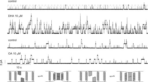
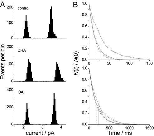
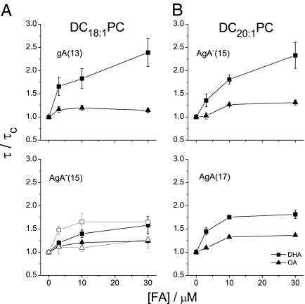
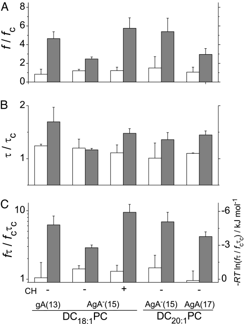
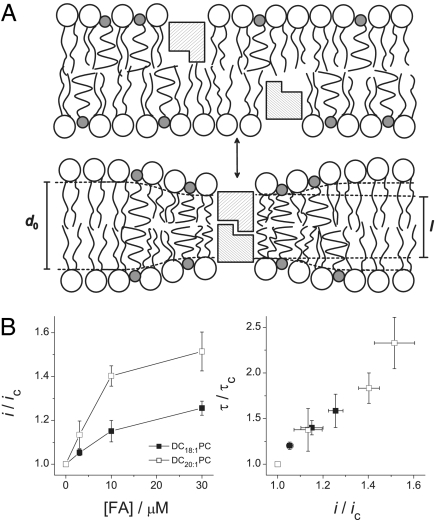
At low micromolar concentrations, polyunsaturated fatty acids (PUFAs) alter the function of many membrane proteins. PUFAs exert their effects on unrelated proteins at similar concentrations, suggesting a common mode of action. Because lipid bilayers serve as the common "solvent" for membrane proteins, the common mechanism could be that PUFAs adsorb to the bilayer/solution interface to promote a negative-going change in lipid intrinsic curvature and, like other reversibly adsorbing amphiphiles, increase bilayer elasticity. PUFA adsorption thus would alter the bilayer deformation energy associated with protein conformational changes involving the protein/bilayer boundary, which would alter protein function. To explore the feasibility of such a mechanism, we used gramicidin (gA) analogues of different lengths together with bilayers of different thicknesses to assess whether docosahexaenoic acid (DHA) could exert its effects through a bilayer-mediated mechanism. Indeed, DHA increases gA channel appearance rates and lifetimes and decreases the free energy of channel formation. The appearance rate and lifetime changes increase with increasing channel-bilayer hydrophobic mismatch and are not related to differing DHA bilayer absorption coefficients. DHA thus alters bilayer elastic properties, not just lipid intrinsic curvature; the elasticity changes are important for DHA's bilayer-modifying actions. Oleic acid (OA), which has little effect on membrane protein function, exerts no such effects despite OA's adsorption coefficient being an order of magnitude greater than DHA's. These results suggest that DHA (and other PUFAs) may modulate membrane protein function by bilayer-mediated mechanisms that do not involve specific protein binding but rather changes in bilayer material properties.


Conflict of interest statement

The authors declare no conflict of interest.

Figures

Fig. 1.
Fig. 1.
Effect of OA and DHA on gA channel activity. Current traces before and after addition of 10 μM DHA (top two traces) or OA (bottom two traces) to both sides of a DC18:1PC/n-decane bilayer containing gA(13) and AgA(15). (Results from two different experiments.) The interrupted lines denote the current levels for gA(13) (short dash) and AgA(15) (long dash). 1 M NaCl, 10 mM Hepes, pH 7, ± 200 mV, 500 Hz. The cartoons at the bottom of the figure illustrate the differences in bilayer deformation with differing hydrophobic mismatch between channel (shaded blocks) and lipid bilayer (represented by springs).
Fig. 2.
Fig. 2.
Effect of OA and DHA on gA single-channel current transitions and lifetimes. (A) Current transition amplitude histograms of gA(13) (left peak) and AgA(15) (right peak) channels in DC18:1PC bilayers in the absence or presence of 10 μM DHA or OA. (B) Normalized single-channel survivor histograms for gA(13) (Upper) and AgA(15) (Lower) fitted with single exponential distributions; note the 10-fold difference in the scale of the abscissae. The vertical dotted lines indicate the average channel lifetimes of (from left to right) control, 10 μM OA, and 10 μM DHA.
Fig. 3.
Fig. 3.
Concentration dependence of OA's and DHA's effects on channel lifetimes. Changes in the lifetime relative to control are plotted as normalized dose-response curves for DHA (filled square) and OA (filled triangle). (A) gA(13) and AgA(15) channels in DC18:1PC bilayers, as well as AgA(15) channels in DC18:1PC:Chol (1:1) bilayers [DHA (open squares) and OA (open triangles) in Lower]; (B) AgA(15) and AgA(17) channels in DC20:1PC bilayers.
Fig. 4.
Fig. 4.
Changes in kinetics and energetics of channel formation. (A) The relative increase in channel appearance rate (f) induced by 3 μM OA (white) or DHA (gray) in DC18:1PC, DC18:1PC:Chol (1:1) and DC20:1PC bilayers. (B) Corresponding changes in gA channel lifetimes (τ). (C) Changes in dimerization constant (left axis) and free energy of dimerization (right axis) for gA channel formation induced by 3 μM OA or DHA. τc, lifetime in the absence of fatty acid; τ, lifetime in the presence of fatty acid; fc, channel appearance rate in the absence of fatty acid; f, appearance rate in the presence of fatty acid.
Fig. 5.
Fig. 5.
DHA alteration of bilayer mechanical properties. (A) Schematic model in which DHA is enriched around the channel in the area of bilayer deformation. l is the channel hydrophobic length and d0 the bilayer hydrophobic thickness. (B) Changes in AgA(15) channel properties by DHA in DC18:1PC (filled squares) and DC20:1PC (open squares). (Left) Normalized single-channel current (i) changes. (Right) Changes in τ as a function of changes in i.

References

    1. Bucher HC, Hengstler P, Schindler C, Meier G. Am J Med. 2002;112:298–304. - PubMed
    1. Kang JX. J Membr Biol. 2005;206:165–172. - PubMed
    1. Hooper L, Thompson RL, Harrison RA, Summerbell CD, Ness AR, Moore HJ, Worthington HV, Durrington PN, Higgins JP, et al. BMJ. 2006;332:752–760. - PMC - PubMed
    1. Leaf A, Xiao YF. J Membr Biol. 2001;184:263–271. - PubMed
    1. McLennan PL, Abeywardena MY, Charnock JS. Can J Physiol Pharmacol. 1985;63:1411–1417. - PubMed

Publication types

MeSH terms

LinkOut - more resources