. 2007 Jun 12;104(24):10051-6.
doi: 10.1073/pnas.0700007104. Epub 2007 May 29.

Inhibition of ubiquitin-mediated degradation of MOAP-1 by apoptotic stimuli promotes Bax function in mitochondria

Affiliations

Inhibition of ubiquitin-mediated degradation of MOAP-1 by apoptotic stimuli promotes Bax function in mitochondria

Nai Yang Fu et al. Proc Natl Acad Sci U S A. .

Abstract

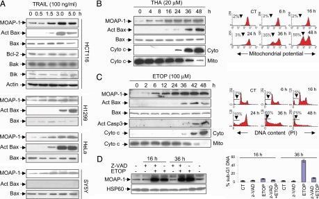
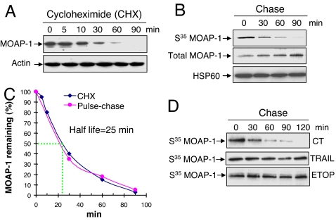
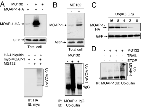
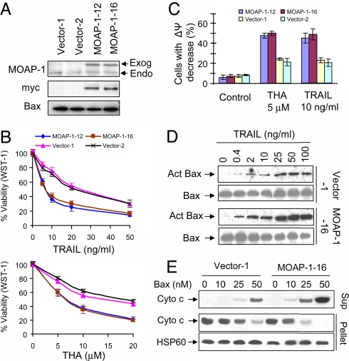
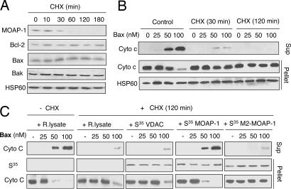
The multidomain proapoptotic protein Bax of the Bcl-2 family is a central regulator for controlling the release of apoptogenic factors from mitochondria. Recent evidence suggests that the Bax-associating protein MOAP-1 may act as an effector for promoting Bax function in mitochondria. Here, we report that MOAP-1 protein is rapidly up-regulated by multiple apoptotic stimuli in mammalian cells. MOAP-1 is a short-lived protein (t(1/2) approximately 25 min) that is constitutively degraded by the ubiquitin-proteasome system. Induction of MOAP-1 by apoptotic stimuli ensues through inhibition of its polyubiquitination process. Elevation of MOAP-1 levels sensitizes cells to apoptotic stimuli and promotes recombinant Bax-mediated cytochrome c release from isolated mitochondria. Mitochondria depleted of short-lived proteins by cycloheximide (CHX) become resistant to Bax-mediated cytochrome c release. Remarkably, incubation of these mitochondria with in vitro-translated MOAP-1 effectively restores the cytochrome c releasing effect of recombinant Bax. We propose that apoptotic stimuli can facilitate the proapoptotic function of Bax in mitochondria through stabilization of MOAP-1.


Conflict of interest statement

The authors declare no conflict of interest.

Figures

Fig. 1.
Fig. 1.
Apoptotic stimuli up-regulate MOAP-1 protein during the early phase of apoptotic signaling. (A) Levels of endogenous MOAP-1 protein were rapidly up-regulated by TRAIL. The indicated cells were treated with TRAIL for various periods of time. RIPA lysates were subjected to IP with the rabbit anti-MOAP-1 antibody (R5), followed by IB with the mouse anti-MOAP-1 antibody (M6). Actin was used as an internal control to demonstrate that equal amount of total proteins was used for IP. The levels of Bcl-2, Bak, and Bik in the total lysates of HCT116 cells were also measured with their respective antibodies. Bax activation (Act Bax) was monitored by using a conformation-specific Bax antibody (N-20). (B and C) Induction of MOAP-1 by THA and ETOP occurred during the early phase of apoptotic signaling. HCT116 cells (B) or 293T cells (C) were treated with 20 μM THA or 100 μM ETOP, respectively, for the indicated periods of time. MOAP-1 levels, Bax activation (Act Bax), Capase 3 activation (Act Casp3), and Cyto c release were monitored (B and C Left). For detection of Cyto c release from mitochondria, the cells were fractionated into heavy membrane fractions enriched with mitochondria (Mito) and cytosolic (Cyto) fractions. THA-induced mitochondrial depolarization (B Right) or ETOP-induced DNA fragmentation (C Right) were analyzed by flow cytometry as described in Materials and Methods. Data shown are representative of at least three independent experiments. (D) Caspase inhibition fails to suppress the elevation of MOAP-1 protein induced by ETOP. 293 T cells were pretreated with 10 μM z-VAD for 1 h before being subjected to 100 μM ETOP treatment for 16 h or 36 h. (Left) MOAP-1 levels were analyzed as in A. (Right) DNA fragmentation was analyzed as in C. Results are presented as percentage of apoptotic cells (mean ± SD, n = 3).
Fig. 2.
Fig. 2.
MOAP-1 is a short-lived protein that can be stabilized by apoptotic stimuli. (A) Inhibition of de novo protein synthesis caused rapid elimination of endogenous MOAP-1 protein. 293T cells were incubated with 50 μg/ml CHX for the indicated periods of time. MOAP-1 protein levels were monitored as in Fig. 1. Actin was used as an internal control. (B) Pulse–chase analysis for estimating the half-life of MOAP-1. 293T cells labeled with S35-methionine/cysteine were chased for the indicated periods of time. RIPA lysates were subjected to IP with anti-MOAP-1 antibody (R5). Immunoprecipitates were analyzed by autoradiography (for S35 MOAP-1) or by IB with anti-MOAP-1 antibody (for total MOAP-1). HSP60 was used as an internal control. (C) The half-life of MOAP-1 is estimated to be ≈25 min. The relative amount of total MOAP-1 in A or S35-labeled MOAP-1 in B was quantified by densitometry and plotted with respect to time. MOAP-1 level at time 0 was defined as 100%. (D) TRAIL or ETOP treatment extended the half-life of MOAP-1. H1299 cells were pretreated with 50 ng/ml TRAIL for 1 h or 100 μM ETOP for 3 h. Pulse–chase assay was then performed as in B. The same concentration of TRAIL or ETOP was maintained during the entire period of pulse–chase analysis.
Fig. 3.
Fig. 3.
Apoptotic stimuli suppress polyubiquitination of MOAP-1. (A) Proteasome inhibition caused accumulation of polyubiquitinated forms of transiently expressed MOAP-1 (Ub MOAP-1). (Upper) HA-MOAP-1 or control vector was cotransfected with pEGFP into 293T cells. Sixteen hours after transfection, the cells were treated with 10 μM MG132 for another sixteen hours. RIPA lysates were analyzed by IB, using anti-HA antibody. The levels of GFP were used to monitor transfection efficiency. (Lower) 293T cells were transfected with indicated plasmids. Sixteen hours after transfection, the cells were left untreated or treated with MG132 for another 16 h. RIPA lysates were subjected to IP with anti-HA-conjugated beads, followed by IB with anti-myc antibody. (B) Proteasome inhibition caused accumulation of ubiquitinated forms of endogenous MOAP-1 (Ub MOAP-1). SY5Y cells were either left untreated or treated with 10 μM MG132 for 16 h. (Upper) RIPA lysates were analyzed by IB with anti-MOAP-1 or anti-actin (loading control) antibodies. (Lower) RIPA lysates were subjected to IP with MOAP-1 antibody or control IgG, followed by IB with anti-Ub antibody. (C) MOAP-1 protein levels were elevated by inhibition of polyubiquitin chain formation. 293T cells were cotransfected with MOAP-1-HA and pEGFP in combination with indicated amounts of plasmid expressing the lysine-less Ub mutant [Ub(K0)]. Sixteen hours after transfection, the cells were harvested, and RIPA lysates were analyzed by IB with anti-HA antibody. The levels of GFP were used to monitor transfection efficiency. (D) TRAIL and ETOP inhibit polyubiquitination of MOAP-1. H1299 cells were treated with 50 ng/ml TRAIL for 1 h or 100 μM ETOP for 3 h before MG132 was added, and the cells were incubated for another 12 h. The cells were harvested and analyzed as in B.
Fig. 4.
Fig. 4.
Higher levels of MOAP-1 sensitize the HCT116 cells to multiple apoptotic stimuli. (A) MOAP-1 expression in the HCT116 vector control and myc-MOAP-1 stable clonal lines. RIPA lysates were immunoprecipitated with anti-MOAP-1 or anti-Bax antibodies, followed by IB with anti-MOAP-1, anti-Bax or anti-myc antibodies. The arrows indicate the endogenous (Endo) or exogenous myc-MOAP-1(Exog). (B and C) Cell death analysis of HCT116 vector control or HCT116 myc-MOAP-1 cells subjected to treatment with TRAIL or THA. Cells grown in 96-well plate were treated with indicated concentrations of THA for 36 h or TRAIL for 16 h. Cell viability after treatment was determined by the WST-1 assay (B). Results are presented as percentage of control (mean ± SD, n = 3). Cells grown in 6-well plate were treated with 5 μM THA for 36 h or 10 ng/ml TRAIL for 16 h, harvested and stained with Mito-tracker Red for analysis of mitochondrial membrane potential changes by flow cytometry (C). (D) Higher levels of MOAP-1 promote TRAIL-induced Bax activation. Vector 1# and MOAP-1 16# cells were treated with indicated concentrations of TRAIL for 5 h. Bax activation (Act Bax) was analyzed by using a conformation-specific Bax antibody (N-20). (E) Higher levels of MOAP-1 increase the sensitivity of isolated mitochondria to recombinant Bax-induced Cyto c release. Heavy membrane fractions containing mitochondria isolated from Vector-1 or MOAP-1–16 cells were treated with recombinant Bax, followed by centrifugation. The supernatants (sup) and pellets were immunoblotted with anti-Cyto c or HSP60 antibodies.
Fig. 5.
Fig. 5.
MOAP-1 is a key short-lived protein to facilitate Bax-induced Cyto c release from isolated mitochondria. (A) Effects of CHX on the levels of Bcl-2 family proteins in heavy membrane fractions containing mitochondria. HCT116 cells were treated with 50 μg/ml CHX for the indicated periods of time. The levels of MOAP-1, Bcl-2, Bak, Bax, and HSP60 were analyzed by IB. (B) Mitochondria isolated from CHX-pretreated cells were resistant to Bax-mediated Cyto c release. Heavy membrane fractions containing mitochondria isolated from the control or CHX-pretreated HCT116 cells were incubated with recombinant Bax followed by centrifugation. The supernatants (sup) and pellets were immunoblotted with anti-Cyto c or HSP60 antibodies. (C) In vitro-translated MOAP-1 protein effectively restored the sensitivity of mitochondria from CHX-pretreated cells to the Cyto c releasing effect of Bax. Heavy membrane fractions containing mitochondria from the cells pretreated with CHX for 2 h were preincubated with PBS, rabbit reticulate lysate (R. lysate), S35-labeled (1 × 10⁴ cpm/methionine) in vitro-translated MOAP-1, VDAC, or the M2 mutant of MOAP-1 (amino acids 1–115) for 20 min. The mitochondria was washed twice, treated with recombinant Bax, and analyzed as in B.

References

    1. Wang X. Genes Dev. 2001;15:2922–2933. - PubMed
    1. Chan SL, Yu VC. Clin Exp Pharmacol Physiol. 2004;31:119–128. - PubMed
    1. Danial NN, Korsmeyer SJ. Cell. 2004;116:205–219. - PubMed
    1. Strasser A. Nat Rev Immunol. 2005;5:189–200. - PubMed
    1. Willis SN, Adams JM. Curr Opin Cell Biol. 2005;17:617–625. - PMC - PubMed

Publication types

MeSH terms