. 2007 Jun 12;104(24):10217-22.
doi: 10.1073/pnas.0608585104. Epub 2007 May 29.

Alarin is a vasoactive peptide

Affiliations

Alarin is a vasoactive peptide

Radmila Santic et al. Proc Natl Acad Sci U S A. .

Abstract

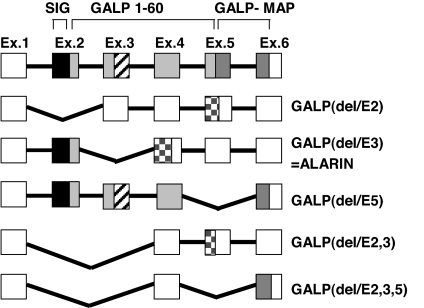
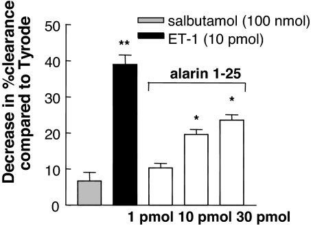
Galanin-like peptide (GALP) is a hypothalamic neuropeptide belonging to the galanin family of peptides. The GALP gene is characterized by extensive differential splicing in a variety of murine tissues. One splice variant excludes exon 3 and results in a frame shift leading to a novel peptide sequence and a stop codon after 49 aa. In this peptide, which we termed alarin, the signal sequence of the GALP precursor peptide and the first 5 aa of the mature GALP are followed by 20 aa without homology to any other murine protein. Alarin mRNA was detected in murine brain, thymus, and skin. In accordance with its vascular localization, the peptide exhibited potent and dose-dependent vasoconstrictor and anti-edema activity in the cutaneous microvasculature, as was also observed with other members of the galanin peptide family. However, in contrast to galanin peptides in general, the physiological effects of alarin do not appear to be mediated via the known galanin receptors. Alarin adds another facet to the surprisingly high-functional redundancy of the galanin family of peptides.


Conflict of interest statement

The authors declare no conflict of interest.

Figures

Fig. 1.
Fig. 1.
Murine prepro-GALP gene splice variants. Filled boxes indicate translated peptides. Open boxes indicate 3′ and 5′ untranslated mRNA regions. Black boxes represent the signal peptide (SIG). Light gray squares indicate the mature GALP peptide with homology to galanin (cross-striped boxes). Dark gray boxes represent the GALP message-associated peptide (GALP-MAP), and checkered boxes represent the putative novel peptide sequences. Exon sizes are not drawn to scale. GALP(delE3) was termed alarin.
Fig. 2.
Fig. 2.
Amino acid sequence comparison of murine, rat, macaque, and human alarin. The arrow indicates a putative N-terminal proteolytic cleavage site of dipeptidyl dipeptidase IV. The solid underline indicates the GALP/alarin shared residues.
Fig. 3.
Fig. 3.
Expression of GALP mRNAs in different murine tissues. RT-PCR was carried out by using total RNA extracted from different tissues of normal mice. Specific primer sets, as shown in SI Table 1, were used to amplify specific splice variants. PCR products were visualized by ethidium bromide staining after agarose gel electrophoresis. Lanes 1–6, murine brain; lanes 7–12, murine skin; lanes 13–18, murine thymus. Specific PCR amplifications for full-length GALP (lanes 1, 7, and 13), GALP(del/E2) (lanes 2, 8, and 14), GALP(del/E3) = alarin (lanes 3, 9, and 15), GALP(del/E5) (lanes 4, 10, and 16), GALP(del/E2,3) (lanes 5, 11, and 17), and GALP(del/E2,3,5) (lanes 6, 12, and 18) are shown. Lane M, Alpha Quant 5 DNA Ladder (Alpha Innotech).
Fig. 4.
Fig. 4.
Alarin-LI in the dermal microvasculature. Alarin-LI in paraffin sections of murine (A and C) and human (B, D–G, and I) dermal blood vessels. Immunohistochemistry was performed with affinity-purified anti-murine alarin (6–24) antiserum (A and C) and anti-human alarin (6–24) antiserum (B, D–G, and I). Immunohistochemistry was performed with antisera preabsorbed with 3 μM synthetic murine (C) and human (D) alarin 1–25 peptide. Alarin-LI in precapillary arterioles (E) and postcapillary venules of different size (F, G, and I) are shown. Comparison of alarin-LI (G and I) with actin immunoreactivity (H and J) indicates staining of alarin-LI in perivascular cell types (pericytes and smooth muscle cells) but not endothelial cells of human microvessels. I and J represent higher magnification of the boxed area in G and H, respectively. Arrows point toward vascular endothelial cells. Green staining indicates alarin-LI and actin immunoreactivity. (Scale bars: 50 μm.)
Fig. 5.
Fig. 5.
Effects on inflammatory edema of alarin. (A) Edema formation was induced by intradermal SP (300 pmol) and CGRP (10 pmol), and the effect of coinjected alarin and different galanin peptides is shown (n = 6). The response of increasing doses of alarin (1–25) (B) and alarin (3–25) (C) coinjected with SP plus CGRP is shown (n = 8). Results are expressed as plasma extravasation (μl/g), mean ± SEM, measured by the 125I-BSA method. Responses that are significantly different from the corresponding SP plus CGRP-treated sites are indicated (∗, P < 0.05; ∗∗, P < 0.01).
Fig. 6.
Fig. 6.
The dose-related effect of alarin on blood flow in cutaneous dorsal microvasculature. The responses of salbutamol and endothelin-1 (ET-1) as controls are shown alongside responses for alarin 1–25. The dose–response effect is shown as a decrease in percentage clearance (mean ± SEM, n = 8) compared with vehicle (Tyrode-injected) skin. Results that are significantly different from Tyrode-injected sites are indicated (∗, P < 0.05; ∗∗, P < 0.01).

Similar articles

Cited by

References

    1. Hoyle CH. Brain Res. 1999;848:1–25. - PubMed
    1. Tatemoto K, Rokaeus A, Jornvall H, McDonald TJ, Mutt V. FEBS Lett. 1983;164:124–128. - PubMed
    1. Ohtaki T, Kumano S, Ishibashi Y, Ogi K, Matsui H, Harada M, Kitada C, Kurokawa T, Onda H, Fujino M. J Biol Chem. 1999;274:37041–37045. - PubMed
    1. Krasnow SM, Fraley GS, Schuh SM, Baumgartner JW, Clifton DK, Steiner RA. Endocrinology. 2003;144:813–822. - PubMed
    1. Gottsch ML, Clifton DK, Steiner RA. Trends Endocrinol Metab. 2004;15:215–221. - PubMed

Publication types

MeSH terms

Associated data

LinkOut - more resources