Comparative Study
. 2007 Jun 5;104(23):9870-5.
doi: 10.1073/pnas.0700436104. Epub 2007 May 29.

Mechanism of the 5-hydroxytryptamine 2A receptor-mediated facilitation of synaptic activity in prefrontal cortex

Affiliations
Comparative Study

Mechanism of the 5-hydroxytryptamine 2A receptor-mediated facilitation of synaptic activity in prefrontal cortex

Jean-Claude Béïque et al. Proc Natl Acad Sci U S A. .

Abstract

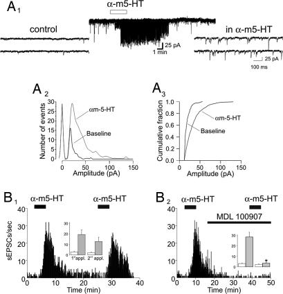
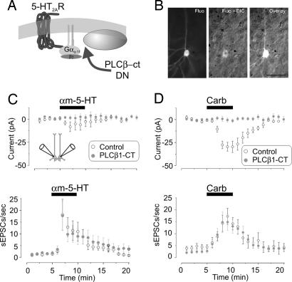
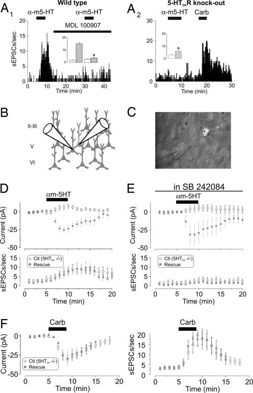
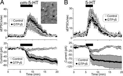
Classic hallucinogens such as lysergic acid diethylamide are thought to elicit their psychotropic actions via serotonin receptors of the 5-hydroxytryptamine 2A subtype (5-HT(2A)R). One likely site for these effects is the prefrontal cortex (PFC). Previous studies have shown that activation of 5-HT(2A)Rs in this region results in a robust increase in spontaneous glutamatergic synaptic activity, and these results have led to the widely held idea that hallucinogens elicit their effect by modulating synaptic transmission within the PFC. Here, we combine cellular and molecular biological approaches, including single-cell 5-HT(2A)Rs inactivation and 5-HT(2A)R rescue over a 5-HT(2A)R knockout genetic background, to distinguish between competing hypotheses accounting for these effects. The results from these experiments do not support the idea that 5-HT(2A)Rs elicit the release of an excitatory retrograde messenger nor that they activate thalamocortical afferents, the two dominant hypotheses. Rather, they suggest that 5-HT(2A)Rs facilitate intrinsic networks within the PFC. Consistent with this idea, we locate a discrete subpopulation of pyramidal cells that is strongly excited by 5-HT(2A)R activation.


Conflict of interest statement

The authors declare no conflict of interest.

Figures

Fig. 1.
Fig. 1.
5-HT2ARs increase synaptic activity in the PFC. (A1) Administration of αm-5-HT (10 μM) results in a large increase in both the frequency and amplitude of sEPSCs recorded from a layer V pyramidal neuron. (A2) Histogram depicting the effect of αm-5-HT on the distribution of sEPSC amplitudes for the experiment illustrated in A1. The leftmost distribution centered at 0 pA depicts the noise distribution. (A3) Cumulative distribution of sEPSC amplitudes recorded before and after the administration of αm-5-HT for the same experiment. (B1) In a different cell, two consecutive administrations of αm-5-HT (10 μM) result in reliable increases in sEPSC activity. (Inset) Plot summarizing the effect of consecutive applications of αm-5-HT on sEPSCs (n = 6). White bars, baseline; gray bars, αm-5-HT. (B2) Administration of MDL 100907 (300 nM) blocks the ability of αm-5-HT to elicit an increase in sEPSC activity. (Inset) Plot summarizing the effect of MDL 100907 (n = 7). (∗, P < 0.01, paired Student's t test). White bars, baseline; gray bars, αm-5-HT.
Fig. 2.
Fig. 2.
Single-cell inactivation of 5-HT2ARs by PLCβ-ct blocked the 5-HT2AR-induced inward current but not the increase in sEPSCs. (A) Diagram illustrating the site of action for the PLCβ-ct dominant negative. (B) Differential interference contrast (DIC)/fluorescence (Fluo) image of a neuron transfected with the PLCβ-ct dominant negative in an organotypic cortical slice (postnatal day 12; 3 days in vitro). (Scale bar: 50 μm.) (C) Paired recordings from neighboring neurons (Inset; n = 14) showing that the inward current elicited by αm-5-HT in control neurons was blocked in neurons expressing the PLCβ-ct construct (Upper; P < 0.05, paired Student's t test) but not the increase in sEPSCs (Lower; P = 0.59, paired Student's t test; n = 14 pairs). (D) Administration of carbachol (Carb) induced a slow inward current in control, nontransfected neurons (−29.9 ± 6.1 pA; n = 11) but not in neurons transfected with the PLCβ-ct construct (−0.4 ± 2.4 pA; n = 20; P < 0.01, unpaired Student's t test) (Upper). The ability of carbachol to induce an increase in sEPSCs was indistinguishable between control and transfected neurons (Lower; P = 0.41, unpaired Student's t test).
Fig. 3.
Fig. 3.
5-HT2AR expression in cells derived from 5-HT2AR knockout (KO) mice rescues the ability of αm-5-HT to induce an inward current but not to increase sEPSC activity. (A1) Administration of αm-5-HT (10 μM) increases sEPSC activity in wild-type mice, and this effect was blocked by MDL 100907 (300 nM). (Inset) Plot summarizing the effect of this experiment in four neurons (∗, P < 0.01, paired Student's t test). White bars, baseline; gray bars, αm-5-HT. (A2) The ability of αm-5-HT to increase sEPSC activity is blocked in this recording derived from a 5-HT2AR KO mouse. In this same cell, carbachol (Carb) (30 μM) elicited a robust increase in sEPSC activity. On average, the ability of αm-5-HT to increase sEPSCs activity was largely, although not completely, blocked in the KO (Inset) (n = 39; residual increase in sEPCS by αm-5-HT; ∗, P < 0.01 compared with baseline, paired Student's t test). White bars, baseline; gray bars, αm-5-HT. (B) Diagram illustrating the approach used for this experiment. 5-HT2ARs were expressed in cultured cortical brain slices derived from 5-HT2AR KO mice to rescue 5-HT2AR expression in a small subset of neurons. (C) Differential interference contrast/fluorescence image illustrating a paired recording from a control neuron and one transfected with 5-HT2ARs and GFP. (D) Results from paired recordings showing that expression of 5-HT2ARs rescued the ability of αm-5-HT to induce an inward current (Upper; P < 0.01, paired Student's t test; n = 16 pairs) but had no effect on the ability of this agonist to increase sEPSC activity (Lower). (E) In slices bathed in the 5-HT2C receptor antagonist SB 242084 (100 nM; >20 min), paired recordings from control and transfected neurons showed that expression of 5-HT2ARs rescued the ability of αm-5-HT to induce an inward current (Upper; P < 0.01, paired Student's t test; n = 10 pairs) but had no effect on the ability of this agonist to increase sEPSC activity (Lower). (F) The ability of carbachol to induce an inward current (Left) and an increase in sEPSCs (Right) was indistinguishable between controls (n = 16) and neurons transfected with 5-HT2ARs (n = 15) in slices derived from 5-HT2AR KO mice.
Fig. 4.
Fig. 4.
Effects of intracellular GTPγS on the 5-HT2AR-induced inward current and increase in sEPSCs. (A) Cell pairs were patched with electrodes filled with intracellular solution containing either GTP or GTPγS. Intracellular perfusion with GTPγS (>10 min) rendered the inward current induced by αm-5-HT (10 μM) effectively irreversible (Upper) but had no detectable effect on the increase in sEPSCs (n = 13 pairs) (Lower). (Inset) Image illustrating the recording configuration used for this experiment. (B) Essentially identical results were observed in cortical slices derived from adult (postnatal day > 33) rats by using 5-HT as an agonist (10 μM, n = 7 control and n = 6 GTPγS-loaded neurons).
Fig. 5.
Fig. 5.
A subpopulation of pyramidal neurons in acute rat PFC slices is excited by activation of 5-HT2ARs. (A1) Administration of 5-HT (30 μM) depolarized and excited this pyramidal neuron in PFC. (Inset) Image of this cell after filling with biocytin. (Scale bar: 150 μm.) (A2) Administration of αm-5-HT (10 μM) similarly depolarized and excited another pyramidal neuron. (B) Consecutive applications of 5-HT (30 μM) elicited comparable (i.e., nondesensitizing) excitation of this subpopulation of neurons. The excitation induced by 5-HT was blocked by MDL 100907. Upper, n = 6; Lower, n = 7. (C) DAMGO suppresses the 5-HT2AR-induced increase in spontaneous activity. (C1) Consecutive administration of αm-5-HT (30 μM) elicited comparable (i.e., nondesensitizing) increase in frequency of sEPSCs (n = 6). (C2) The increase in sEPSCs induced by αm-5-HT is blocked by administration of DAMGO (10 μM; n = 6; P < 0.01, paired Student's t test). (D) The ability of αm-5-HT (30 μM) to excite pyramidal neurons is blocked by administration of DAMGO (10 μM; n = 5; P < 0.01, paired Student's t test).

Similar articles

Cited by

References

    1. Wooley DW, Shaw E. Proc Natl Acad Sci USA. 1954;40:228–231. - PMC - PubMed
    1. Glennon RA, Titeler M, McKenney JD. Life Sci. 1984;35:2505–2511. - PubMed
    1. Titeler M, Lyon RA, Glennon RA. Psychopharmacology (Berl) 1988;94:213–216. - PubMed
    1. Aghajanian GK, Marek GJ. Neuropsychopharmacology. 1999;21:16S–23S. - PubMed
    1. Nichols DE. Pharmacol Ther. 2004;101:131–181. - PubMed

Publication types

LinkOut - more resources