Comparative Study
. 2007 Jun 5;104(23):9764-9.
doi: 10.1073/pnas.0700240104. Epub 2007 May 29.

Mastermind-1 is required for Notch signal-dependent steps in lymphocyte development in vivo

Affiliations
Comparative Study

Mastermind-1 is required for Notch signal-dependent steps in lymphocyte development in vivo

Toshinao Oyama et al. Proc Natl Acad Sci U S A. .

Abstract

Mastermind (Mam) is one of the elements of Notch signaling, an ancient system that plays a pivotal role in metazoan development. Genetic analyses in Drosophila and Caenorhabditis elegans have shown Mam to be an essential positive regulator of this signaling pathway in these species. Mam proteins bind to and stabilize the DNA-binding complex of the intracellular domains of Notch and CBF-1, Su(H), Lag-1 (CSL) DNA-binding proteins in the nucleus. Mammals have three Mam proteins, which show remarkable similarities in their functions while having an unusual structural diversity. There have also been recent indications that Mam-1 functionally interacts with other transcription factors including p53 tumor suppressor. We herein describe that Mam-1 deficiency in mice abolishes the development of splenic marginal zone B cells, a subset strictly dependent on Notch2, a CSL protein and Delta1 ligand. Mam-1 deficiency also causes a partially impaired development of early thymocytes, while not affecting the generation of definitive hematopoiesis, processes that are dependent on Notch1. We also demonstrate the transcriptional activation of a target promoter by constitutively active forms of Notch to decrease severalfold in cultured Mam-1-deficient cells. These results indicate that Mam-1 is thus required to some extent for Notch-dependent stages in lymphopoiesis, thus supporting the notion that Mam is an essential component of the canonical Notch pathway in mammals.


Conflict of interest statement

The authors declare no conflict of interest.

Figures

Fig. 1.
Fig. 1.
Targeted disruption of the mouse Mam-1 gene. (A) Schematic representations of the wild-type Mam-1 allele around the exon 1, targeting construct, and mutant allele. Probes for the Southern blot analysis are indicated. RI, EcoRI; RV, EcoRV. (B) Southern blot analysis of DNAs isolated from wild-type (+/+), heterozygous (+/−), and homozygous (−/−) mice. The restriction enzymes and probes used are indicated. The sizes of the hybridizing fragments are also indicated.
Fig. 2.
Fig. 2.
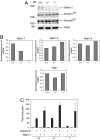
The deficiency of Mam-1 results in a marked decrease of transactivation induced by expression of Notch1IC. (A) A Western blot analysis of the extracts obtained from EFs. Genotypes of the cells, mobility of size markers, and identity of the bands are shown. TM, transmembrane subunit; EC, extracellular subunit. (B) Real-time PCR analysis of RNAs isolated from EFs. Genotypes of the cells and mRNA species analyzed are shown. (C) A luciferase assay with EFs. The cells were transfected with a reporter, an internal control for transfection, and expression vectors for indicated proteins (+) or its empty vectors (−). The vertical axis represents the mean value of normalized relative luciferase activity to the mean activity of the wild-type cells transfected with empty vector controls. The error bars indicate SD (n = 3).
Fig. 3.
Fig. 3.
Growth retardation and early death of Mam-1−/− mice. (A) A Mam-1−/− mouse and its littermate at P12. (B) Growth of Mam-1−/− mice and the littermates. (C) Survival of Mam-1−/− mice. A Kaplan–Meier representation of 10 Mam-1−/− mice.
Fig. 4.
Fig. 4.
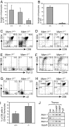
Defective development of early thymocytes in Mam-1−/− mice. (A) The number of thymocytes at E17.5–E18.5. The data are the mean ± SD (n = 5≈18). (B) The number of thymocytes at P7–P8. The data are the mean ± SD (n = 4≈5). (C) A FACS analysis of thymocytes at E17.5. (D) A FACS analysis of thymocytes at P7. (E–G) A FACS analysis of the thymocytes at P7. The profiles were gated on CD4CD8 cells. (H) A FACS analysis of thymocytes from mixed chimeras. The profiles were gated on CD45.2+ cells. The numbers in C–H represent the percentages of cells in the indicated areas. (I) The percentages of DN thymocytes in mixed chimeras. The data are the mean ± SD (n = 3). (J) RT-PCR analysis of mRNAs isolated from fractions of thymocytes. DN, CD4CD8 cells; DP, CD4+CD8+ cells; CD4SP, CD4+CD8 cells; CD8SP, CD4CD8+ cells.
Fig. 5.
Fig. 5.
The defective development of MZB cells from Mam-1−/− hematopoietic stem cells. (A) A FACS analysis of splenic B cells from mixed chimeras. The profiles were gated on CD45.2+B220+ cells. The numbers represent the percentages of the cells in the indicated area (MZB cells). (B) An RT-PCR analysis of mRNAs isolated from fractions of splenic B cells. NFB, newly formed B cells; FOB, follicular B cells.

Similar articles

Cited by

References

    1. Artavanis-Tsakonas S, Rand MD, Lake RJ. Science. 1999;284:770–776. - PubMed
    1. Weng AP, Ferrando AA, Lee W, Morris JP, IV, Silverman LB, Sanchez-Irizarry C, Blacklow SC, Look AT, Aster JC. Science. 2004;306:269–271. - PubMed
    1. Garg V, Muth AN, Ransom JF, Schluterman MK, Barnes R, King IN, Grossfeld PD, Srivastava D. Nature. 2005;437:270–274. - PubMed
    1. Mumm JS, Kopan R. Dev Biol. 2000;228:151–165. - PubMed
    1. Kumano K, Chiba S, Kunisato A, Sata M, Saito T, Nakagami-Yamaguchi E, Yamaguchi T, Masuda S, Shimizu K, Takahashi T, et al. Immunity. 2003;18:699–711. - PubMed

Publication types

LinkOut - more resources