. 2007 Sep 15;110(6):2041-8.
doi: 10.1182/blood-2007-04-082495. Epub 2007 May 29.

The syndecan-1 heparan sulfate proteoglycan is a viable target for myeloma therapy

Affiliations

The syndecan-1 heparan sulfate proteoglycan is a viable target for myeloma therapy

Yang Yang et al. Blood. .

Abstract

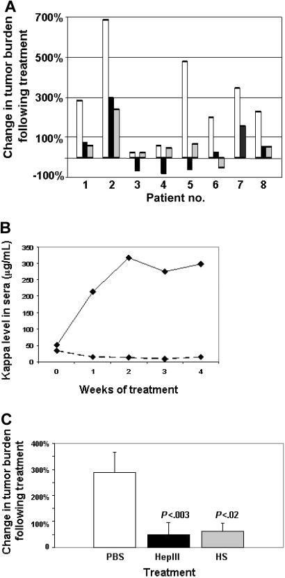
The heparan sulfate proteoglycan syndecan-1 is expressed by myeloma cells and shed into the myeloma microenvironment. High levels of shed syndecan-1 in myeloma patient sera correlate with poor prognosis and studies in animal models indicate that shed syndecan-1 is a potent stimulator of myeloma tumor growth and metastasis. Overexpression of extracellular endosulfatases, enzymes which remove 6-O sulfate groups from heparan sulfate chains, diminishes myeloma tumor growth in vivo. Together, these findings identify syndecan-1 as a potential target for myeloma therapy. Here, 3 different strategies were tested in animal models of myeloma with the following results: (1) treatment with bacterial heparinase III, an enzyme that degrades heparan sulfate chains, dramatically inhibited the growth of primary tumors in the human severe combined immunodeficient (SCID-hu) model of myeloma; (2) treatment with an inhibitor of human heparanase, an enzyme that synergizes with syndecan-1 in promoting myeloma progression, blocked the growth of myeloma in vivo; and (3) knockdown of syndecan-1 expression by RNAi diminished and delayed myeloma tumor development in vivo. These results confirm the importance of syndecan-1 in myeloma pathobiology and provide strong evidence that disruption of the normal function or amount of syndecan-1 or its heparan sulfate chains is a valid therapeutic approach for this cancer.


Figures

Figure 1
Figure 1
HepIII inhibits growth of primary myeloma tumors in vivo. (A) Tumors formed by cells from patients with myeloma were established in the SCID-hu host and then treated for 28 days (patients 1, 2, 4, and 5), 21 days (patients 6-8), or 14 days (patient 3) by daily injection of either PBS (□), active recombinant HepIII enzyme (■), or HepIII-generated heparan sulfate fragments (▩). At the end of the treatment period, human light chain levels in the serum of the mice were analyzed and plotted as the percentage increase or decrease over the light chain level present at the time treatment was initiated. (B) Levels of kappa light chain measured at weekly intervals in animals bearing tumor from patient 5 during treatment with PBS (–) or HepIII (-------). (C) Percentage change in tumor burden following treatment as measured by levels of human light chain in the serum of mice. Bars show the combined mean percentage change of all 8 patients plus or minus SEM following treatment with PBS, HepIII, or heparan sulfate (HS) fragments generated ex vivo by HepIII.
Figure 2
Figure 2
Treatment with HepIII protects implanted bones from osteolysis. At the termination of the experiment, implanted human bones were excised and imaged by microcomputed tomography (microCT). Shown are the three-dimensional reconstructions of the bones (sliced longitudinally through the midpoint of each specimen) injected with cells from patient 4 followed by treatment of the animal with either PBS (excessive bone resorption is seen) or HepIII (no bone resorption is observed, trabecular bone is intact). The areas of increased osteoclastic bone resorption in the PBS-treated sample are indicated by arrowheads. Note the appearance of the blue background behind the bone from the PBS-treated sample, indicative of resorption through the entire specimen. Scale bar equals 1 mm.
Figure 3
Figure 3
An inhibitor of heparanase activity inhibits myeloma tumor growth in vivo. Established subcutaneous tumors formed by CAG myeloma cells expressing heparanase at either high (HPSEhigh) or low (HPSElow) levels were treated for 28 days with PBS or 100NA,RO-H at doses of 18 mg/kg per day (both HPSEhigh and HPSElow cells) or 36 mg/kg per day (HPSEhigh cells only). At termination of the treatment period, tumors were harvested and photographed. X indicates no gross tumor detectable in the animal. Tumor wet weights and P values are shown in Tables 1 and 2.
Figure 4
Figure 4
shRNA reduces syndecan-1 expression on myeloma cells. (A) Expression of syndecan-1 mRNA by lentiviral-infected CAG cells was quantified by PCR (lane 1, control shRNA cells; lane 2, syndecan-1 shRNA cells). Bottom panel shows GAPDH control. Bands migrated to their predicted location relative to standards with sizes of 396 bp for syndecan-1 and 452 bp for GAPDH, respectively. (B) Flow cytometry showing cell-surface expression of syndecan-1 on shRNA control (open peak) or shRNA syndecan-1 (shaded peak) cells. (C) Western blot for syndecan-1 from CAG wild-type (lane 1), control shRNA (lane 2), and syndecan-1 shRNA cells (lane 3).
Figure 5
Figure 5
shRNA inhibits and delays development of myeloma tumors. CAG cells infected with control shRNA (control) or syndecan-1 shRNA (Synlow) were injected subcutaneously into mice. Animals were considered positive for tumor formation when serum kappa light chain levels reached 50 ng/mL. The plot shows the percentage of animals that were free of tumor at weekly intervals for 8 weeks after injection of tumor cells.

Similar articles

Cited by

References

    1. Ridley RC, Xiao HQ, Hata H, Woodliff J, Epstein J, Sanderson RD. Expression of syndecan regulates human myeloma plasma cell adhesion to type I collagen. Blood. 1993;81:767–774. - PubMed
    1. Wijdenes J, Vooijs WC, Clement C, et al. A plasmocyte selective monoclonal antibody (B-B4) recognizes syndecan-1. Br J Haematol. 1996;94:318–323. - PubMed
    1. Bayer-Garner IB, Sanderson RD, Dhodapkar MV, Owens RB, Wilson CS. Syndecan-1 (CD138) immunoreactivity in bone marrow biopsies of multiple myeloma: shed syndecan-1 accumulates in fibrotic regions. Mod Pathol. 2001;14:1052–1058. - PubMed
    1. Dhodapkar MV, Kelly T, Theus A, Athota AB, Barlogie B, Sanderson RD. Elevated levels of shed syndecan-1 correlate with tumour mass and decreased matrix metalloproteinase-9 activity in the serum of patients with multiple myeloma. Br J Haematol. 1997;99:368–371. - PubMed
    1. Seidel C, Sundan A, Hjorth M, et al. Serum syndecan-1: a new independent prognostic marker in multiple myeloma. Blood. 2000;95:388–392. - PubMed

Publication types

MeSH terms