. 2007 May 30:3:11.
doi: 10.1186/1746-6148-3-11.

In vitro binding and survival assays of Leishmania parasites to peripherical blood monocytes and monocyte-derived macrophages isolated from dogs naturally and experimentally infected with Leishmania (Leishmania) chagasi

Affiliations

In vitro binding and survival assays of Leishmania parasites to peripherical blood monocytes and monocyte-derived macrophages isolated from dogs naturally and experimentally infected with Leishmania (Leishmania) chagasi

Weverton M Sampaio et al. BMC Vet Res. .

Abstract

Background: There are a few works considering the characterization of canine monocyte-derived macrophages as well as a standardized procedure for isolation, culture, and infection of these cells with Leishmania. We have performed several modifications in order to improve the canine monocyte-derived macrophage cultures. In addition, we have done a comparative study between monocytes and monocyte-derived macrophages from dogs naturally and experimentally infected with L. chagasi.

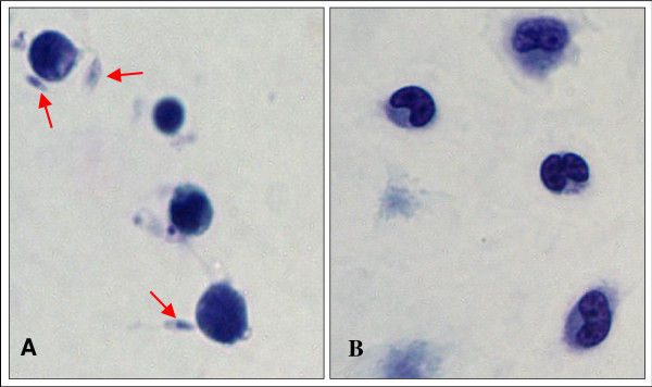
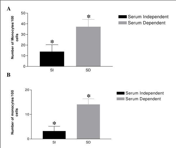
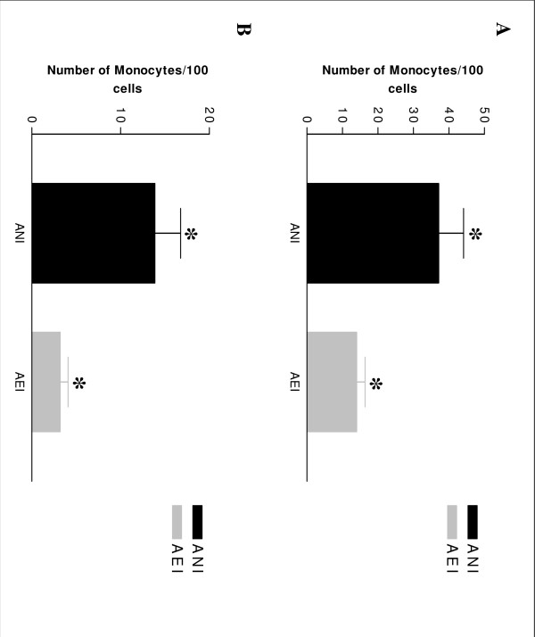
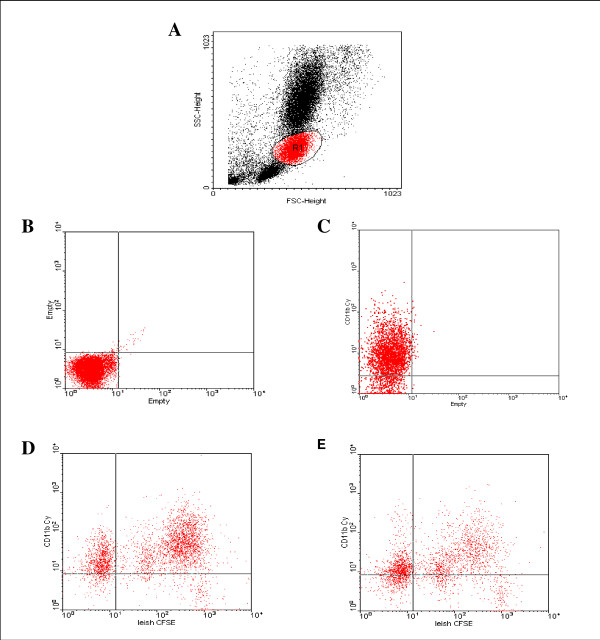
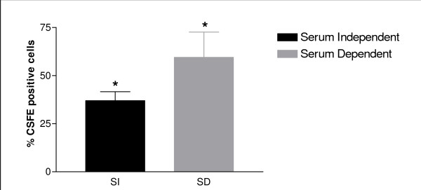
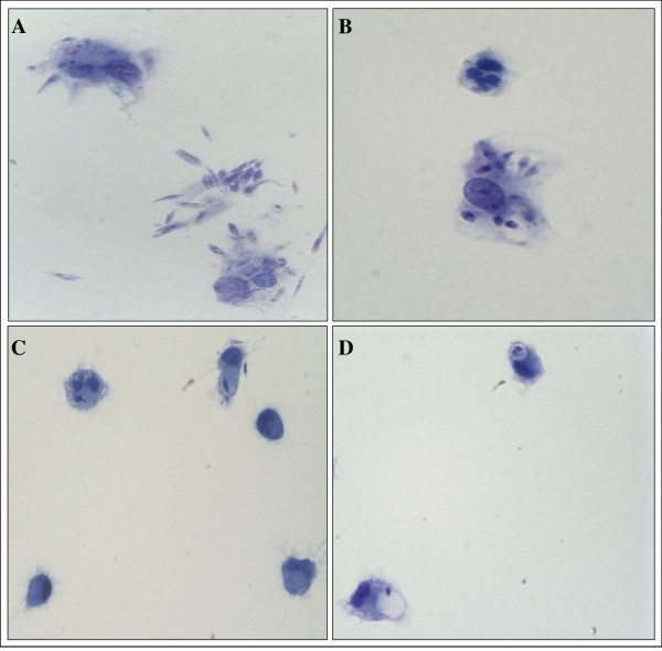
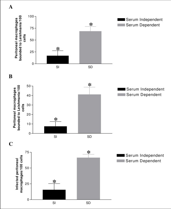
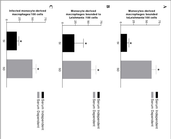
Results: In the presence of exogenous serum, opsonized Leishmania promastigotes binds better to monocytes/macrophages than without serum. Otherwise, this binding occurs due to the strict correlation between the opsonized biologic particles with the third receptor of the complement (CR3-CD11b/CD18). In fact, our assays with CD11b confirmed the importance of this receptor for canine cells and the L. chagasi experimental system. Moreover, monocytes obtained from naturally infected dogs have shown a higher number of monocytes bounded to promastigotes. The experimental results regarding survival have shown that promastigote forms of opsonized L. chagasi were more infective, because we found higher numbers of promastigotes bound to the different cells. As a consequence, after forty-eight hours of binding, higher numbers of amastigotes appeared inside monocyte-macrophages.

Conclusion: These studies have given support to continue comparative studies involving canine monocytes, monocyte-derived macrophages and peritoneal macrophages. Since we have standardized the canine cell culture, we are looking forward to determining the phenotypic properties of these cells before and after L. chagasi infection using flow cytometry.


Figures

Figure 1
Figure 1
The binding of Leishmania promastigotes to canine monocytes. (A) Promastigotes binding to peripherical monocytes from one animal naturally infected (ANI) with Leishmania chagasi in the presence of C5D serum. (arrows) (B) Only monocytes can be observed over coverslips (assay with absence of serum C5D). Giemsa. 1000×.
Figure 2
Figure 2
The binding of Leishmania promastigotes to canine monocytes. (A) Average of Leishmania promastigotes bound to monocytes of animals naturally infected (ANI) with L. chagasi in the presence or absence of C5D serum (2.5 × 10⁶ parasites/well) (* p < 0.01) (B) Average of Leishmania promastigotes bound to monocytes of animals experimentally infected (AEI) with L. chagasi in the presence or absence of C5D serum (2,5 × 10⁶ parasites/well) (*p < 0.01).
Figure 3
Figure 3
The binding of Leishmania promastigotes to canine monocytes. (A) to Leishmania promastigotes bound to monocytes from animals naturally infected (ANI) with L. chagasi and animals experimentally infected (AEI) with L. chagasi in the presence of C5D serum (2.5 × 10⁶ parasites/well) (*) p < 0.01. (B) Leishmania promastigotes bound to monocytes from dogs naturally and experimental infected with L. chagasi in the absence of C5D serum (2.5 × 10⁶ parasites/well) (*) p < 0.01.
Figure 4
Figure 4
Flow cytometer analysis of CD11b integrin expression and the binding of Leishmania promastigotes to canine monocytes. (A) Gate R1 was built separating monocytes from animals naturally infected (ANI) with L. chagasi by cells size (x axis) and granularity (y axis). (B) Gate with mononuclear cells (control) (C) CD11b positive cells; (D) CD11b expression during the L. chagasi binding to mononuclear cells with the presence of C5D serum. (E) CD11b expression during the L. chagasi binding to mononuclear cells with the absence of C5D serum.
Figure 5
Figure 5
Flow cytometer analysis of the binding of Leishmania promastigotes to canine monocytes. Serum dependent conditions induce a higher association of Leishmania with monocytes as measured by both mean fluorescent intensity (CFSE) and percentage of monocytes associated with Leishmania. (*) p < 0.05.
Figure 6
Figure 6
The binding and survival assays of Leishmania promastigotes to monocytes-derived macrophages. (A) Promastigotas adhesion fifty minutes after incubation with the presence of C5D serum. (B) Note many amastigotes into could be seen 48 hours after interaction with the presence of C5D serum. (C) Promastigotas adhesion with the absence of serum. (D) Note some amastigotas could be seen 48 hours after interaction with the absence of C5D serum Giemsa. 1000×.
Figure 7
Figure 7
The binding and survival assays of Leishmania promastigotes to peritoneal macrophages. (A) Average of peritoneal macrophages binding to Leishmania from naturally infected animals with the presence or absence of C5D serum, after 50 min incubation (2,5 × 10⁶ parasites/well) (B) Average of peritoneal macrophages binding to Leishmania from experimental infected animals with the presence or absence of C5D serum, after 50 min incubation (2,5 × 10⁶ parasites/well). (C) Average of infected peritoneal macrophages binding to Leishmania from naturally infected animals with the presence or absence of C5D serum after, fourth eight hour incubation (5 × 10⁶ parasites/well) (*) p < 0.01.
Figure 8
Figure 8
The binding and survival assays of Leishmania promastigotes to monocyte-derived macrophages. (A) Average of monocyte-derived macrophages binding to Leishmania from naturally infected animals with the presence or absence of C5D serum, after 50 min incubation (5 × 10⁶ parasites/well) (B) Average of monocyte-derived macrophages binding to Leishmania from experimental infected animals with the presence or absence of C5D serum, after 50 min incubation (5 × 10⁶ parasites/well). (C) Average of infected peritoneal macrophages binding to Leishmania from experimental infected animals with the presence or absence of C5D serum after, fourth eight hour incubation (5 × 10⁶ parasites/well) (*) p < 0.01.

Similar articles

Cited by

References

    1. Mauricio IL, Stothard JR, Miles MA. The strange case of Leishmania chagasi. Parasitol Today. 2000;16:188–189. doi: 10.1016/S0169-4758(00)01637-9. - DOI - PubMed
    1. Deane LM, Deane MP. Visceral leishmaniasis in Brazil: geographical distribution and trnsmission. Rev Inst Med Trop Sao Paulo. 1962;4:198–212. - PubMed
    1. Pinelli E, Rutten VP, Bruysters M, Moore PF, Ruitenberg EJ. Compensation for decreased expression of B7 molecules on Leishmania infantum-infected canine macrophages results in restoration of parasite-specific T-cell proliferation and gamma interferon production. Infect Immun. 1999;67:237–243. - PMC - PubMed
    1. Shaw SE, Anderson NV. Isolation and functional analysis of normal canine blood monocytes and resident alveolar macrophages. Am J Vet Res. 1984;45:87–90. - PubMed
    1. Tipold A, Zurbriggen A, Moore P, Schijns V, Jungi TW. Generation and functional characterisation of canine bone marrow-derived macrophages. Res Vet Sci. 1998;64:125–132. doi: 10.1016/S0034-5288(98)90007-8. - DOI - PubMed

Publication types