Review
. 2007 Oct 30;136(1-2):1-19.
doi: 10.1016/j.autneu.2007.04.005. Epub 2007 May 29.

Innervation of the gastrointestinal tract: patterns of aging

Affiliations
Review

Innervation of the gastrointestinal tract: patterns of aging

Robert J Phillips et al. Auton Neurosci. .

Abstract

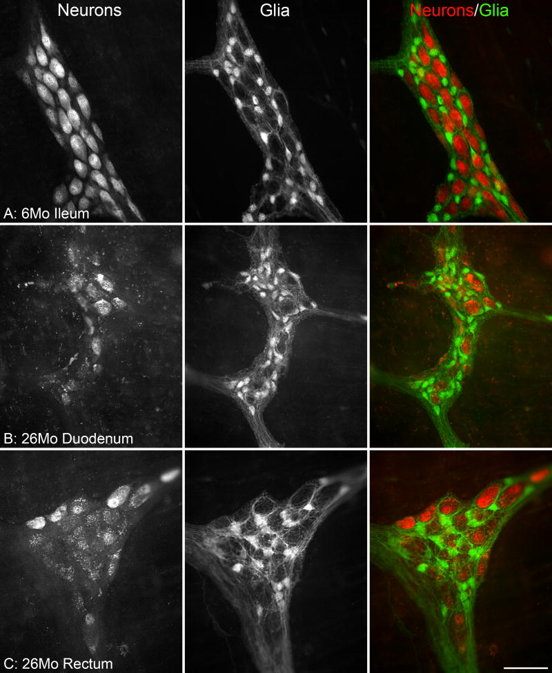
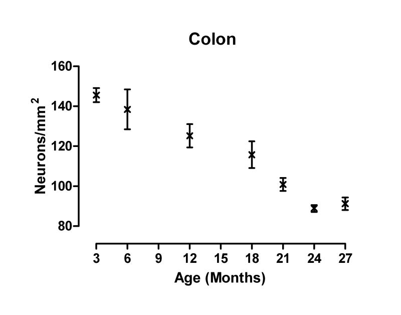
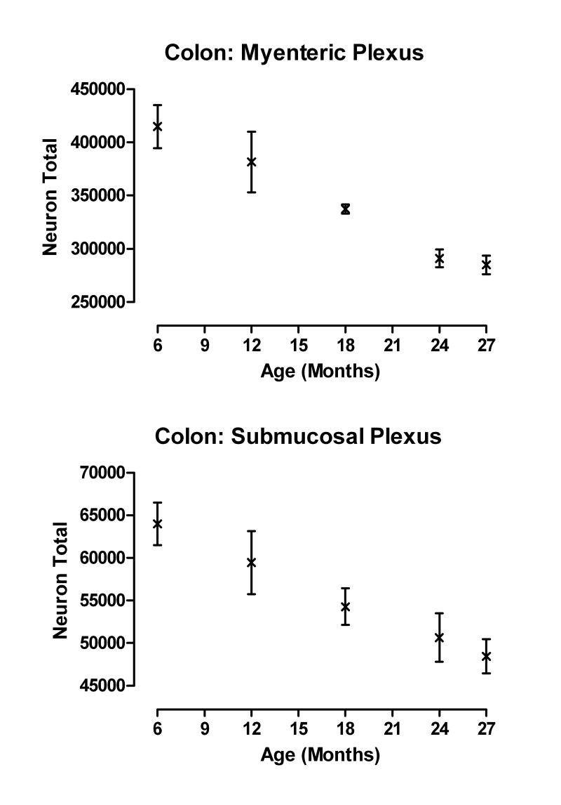
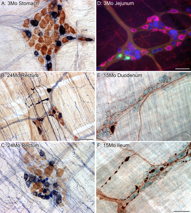
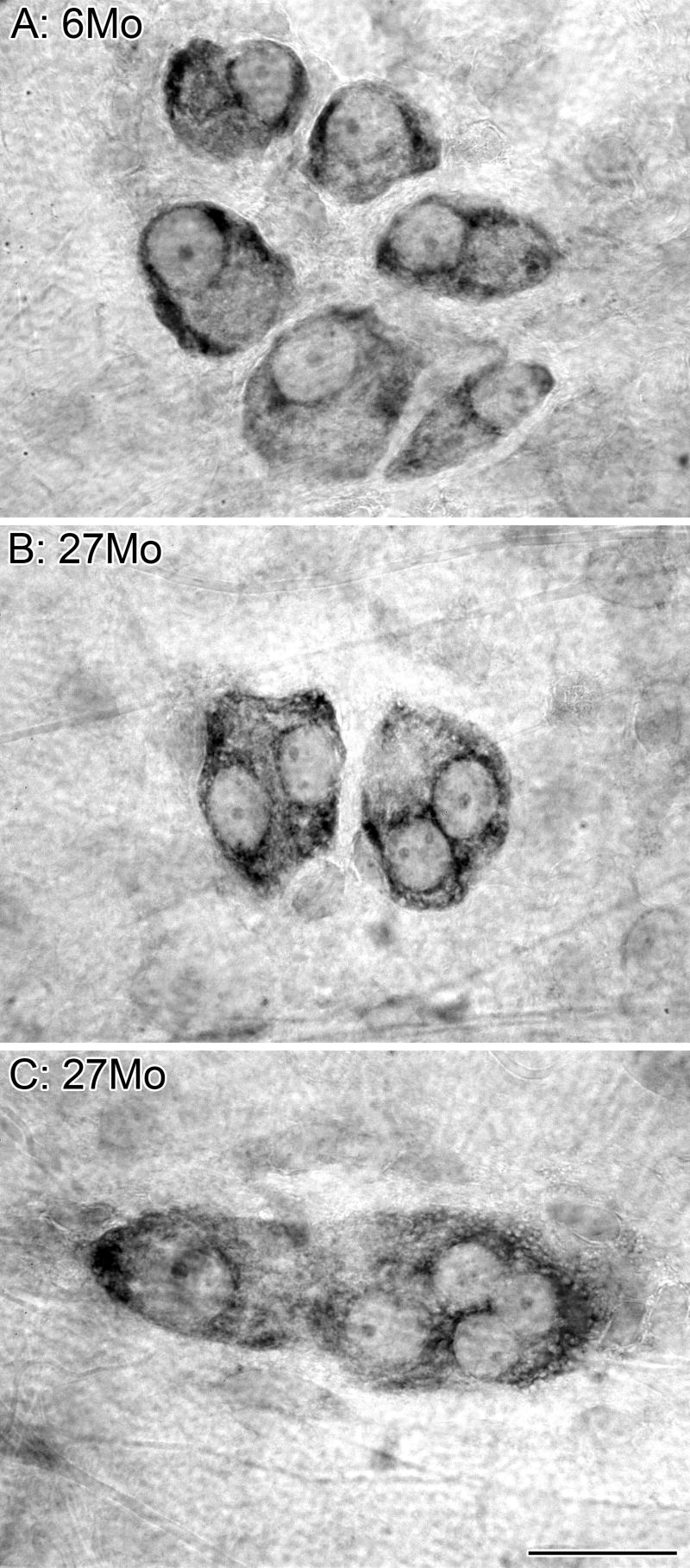
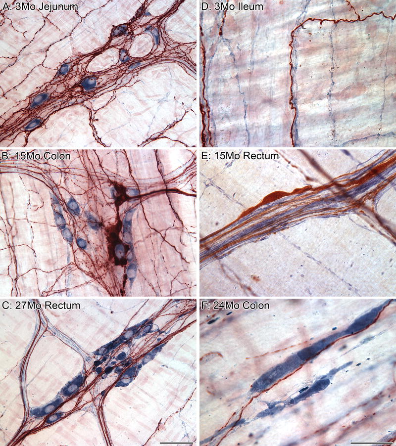
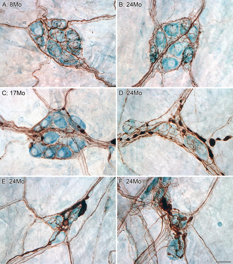
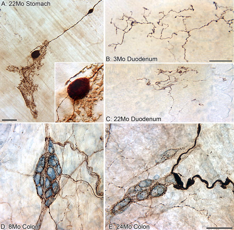
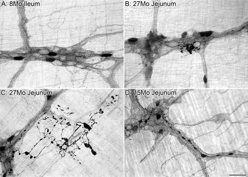
The gastrointestinal (GI) tract is innervated by intrinsic enteric neurons and by extrinsic projections, including sympathetic and parasympathetic efferents as well as visceral afferents, all of which are compromised by age to different degrees. In the present review, we summarize and illustrate key structural changes in the aging innervation of the gut, and suggest a provisional list of the general patterns of aging of the GI innervation. For example, age-related neuronal losses occur in both the myenteric plexus and submucosal plexus of the intestines. These losses start in adulthood, increase over the rest of the life span, and are specific to cholinergic neurons. Parallel losses of enteric glia also occur. The extent of neuronal and glial loss varies along an oral-to-anal gradient, with the more distal GI tract being more severely affected. Additionally, with aging, dystrophic axonal swellings and markedly dilated varicosities progressively accumulate in the sympathetic, vagal, dorsal root, and enteric nitrergic innervation of the gut. These dramatic and consistent patterns of neuropathy that characterize the aging autonomic nervous system of the GI tract are candidate mechanisms for some of the age-related declines in function evidenced in the elderly.


Figures

Figure 1
Figure 1
Dramatic age-related changes occur in the myenteric plexus. Immunostaining of neurons (left column, HuC/D) and glia (middle column, S-100) in the myenteric plexus. The merged images (right column) illustrate the relationship of neurons (red) and glia (green). At 6 months of age, HuC/D strongly labeled the nuclei of neurons with weaker labeling of the cytoplasm and no labeling of the neuronal processes, and S-100 strongly labeled the soma of the glia with weaker labeling of processes (A). At 26 months of age, there was a reduction in the presence of the HuC/D antigen in the neurons of the small (B) and large (C) intestines. The complete loss of HuC/D staining of some neurons at 26 months is in stark contrast to the strong labeling of other neurons within the same ganglion, and is consistent with the observation that age-related neuron loss is specific to a subpopulation of neurons. The complete deterioration of some neurons within a ganglion is indicated by the presence of granular debris or lipofuscin within unstained silhouettes outlined by the glial scaffolding. Scale bar = 50 μm in C (applies to A-C).
Figure 2
Figure 2
Age-related neuronal loss in the myenteric plexus of the colon was evident as early as 12 months of age and progressed in a linear manner with increasing age (combined data from Phillips and Powley, 2001; Phillips et al., 2003, 2006a). A runs test following linear regression analysis verified that the distribution of the data was not significantly nonlinear (runs test, p = 0.54). A mean ± S.E.M of 248 ± 19 neurons/cm2/month are lost with age (goodness of fit, r2 = 0.71). The mean (± S.E.M) density of neurons, which were corrected for age-related changes in tissue area, was 146 (3.6), 138 (10), 125 (5.6), 116 (6.6), 101 (3.3), 89 (1.7), and 91 (3.2) neurons/mm2 at 3, 6, 12, 18, 21, 24, and 27 months of age, respectively. The analysis was based on a sample size of 12, 6, 12, 6, 5, 16, and 12 rats at 3, 6, 12, 18, 21, 24, and 27 months of age, respectively.
Figure 3
Figure 3
The pattern of neuronal loss with age was similar for both the myenteric and submucosal plexuses (data from Phillips et al., 2006a). Total neuronal estimates for both plexuses are from the same rats with 6 rats/age sampled.
Figure 4
Figure 4
The myenteric plexus can be broken down into different subpopulations based upon the neurochemical phenotype of the neurons, and aging appears to affect the subpopulations differently. All of the neurons in the myenteric plexus were labeled using the putative pan-neuronal marker HuC/D (brown), and the subpopulation of neurons that are nitrergic were labeled using the histochemical reaction NADPHd (dark blue; A-C). There is no loss of nitrergic neurons throughout the gastrointestinal tract with age; however, markedly swollen nitrergic axons are routinely found in the smooth muscle (B). In addition, swollen nitrergic axons and dystrophic varicosities were also found in the myenteric ganglia (C; HuC/D neurons brown; nitrergic neurons dark blue with nuclei in silhouette; varicosities solid dark blue elements, some as large as neuronal somata) of aged rats. The majority of the myenteric neurons (blue, HuC/D) in the small intestine of the rat that are positive for the calcium binding protein calbindin (green) are not part of the nitrergic (red, nNOS) phenotype (D). Calbindin-positive (brown) neurons are a small subpopulation of the total population of neurons (light blue, Cuprolinic Blue) within the myenteric plexus (E). We have identified markedly swollen axons in the myenteric plexus of middle-age (15 months of age, F) and aged (24 months of age, not shown) Fischer 344 rats that are positive for calbindin (large brown varicosities and brown axons; blue neuronal counterstain, Cuprolinc Blue). Scale bars = 50 μm in F (applies to A,E,F), D (applies to C,D); 100 μm in B.
Figure 5
Figure 5
A reduction in ganglionic area occurs with age. Cuprolinic Blue labeled neurons in the submucosal plexus of the colon. Neurons were well defined with stained Nissl substance outlining the unstained nuclei. At 6 months of age, neurons within a ganglion were evenly spaced, providing room for unstained axons, dendrites, and glia (A). In the plexus of 27-month-old rats, ganglia were often observed that appeared to have collapsed in upon themselves (B,C), with little or no evidence of neuropil, i.e., the ganglionic tissue surrounding the neuronal cell bodies (e.g., glial cells, nerve fibers and processes of ganglion neurons). Scale bar = 25 μm in C (applies to A-C).
Figure 6
Figure 6
There was no clear relationship between the age-related changes seen in nitrergic and sympathetic neurites. The innervation of nitrergic neurons (dark blue, NADPHd) by sympathetic axons (red-brown, TH) in the ganglia of the myenteric plexus appears, based on the amount of contacts of the varicosities on sympathetic axons with the cytoplasm and nucleus of the nitrergic neurons, to range from sparse to dense (A). In the whole mounts of the myenteric plexus from aging rats, markedly swollen sympathetic axons were found in ganglia with healthy nitrergic innervation (B) and conversely, markedly swollen nitrergic varicosities (dense, dark blue varicosities) were found in ganglia with intact sympathetic innervation (C); however, ganglia with both were never observed in our samples. Sympathetic (red-brown, TH) and nitrergic (dark blue) axons frequently traveled together in the same connectives (D). Markedly swollen sympathetic axons were found coursing through the connective with other sympathetic axons and nitrergic axons of normal diameter (E). Conversely, markedly swollen nitrergic axons in close association with sympathetic axons of normal diameter were frequently found throughout the myenteric plexus (F). Scale bars = 50 μm in C (applies to A-C); 25 μm in F (applies to D-F).
Figure 7
Figure 7
A distinct axonopathy represented by markedly swollen sympathetic axons was commonly found in the innervation of the submucosal plexus. All of the ganglia (blue neuronal counterstain, Cuprolinc Blue) within the submucosal plexus were innervated by sympathetic (brown, TH) axons (A-F). Cases of well-labeled sympathetic axons that were healthy in appearance (i.e., identical to the innervation seen at younger ages; A) were routinely found in aged tissue (B). In contrast, however, swollen sympathetic axons and terminals also became both more common and more pronounced with age (C-F). Ganglia were often observed to be innervated by sympathetic axons that were both markedly swollen and healthy looking (C,D). Scale bar = 25 μm in F (applies to A-F).
Figure 8
Figure 8
Visceral afferent innervation of the gastrointestinal tract evidenced signs of deterioration with age. A parent neurite with two markedly swollen varicosities along its length (brown, labeled using the anterograde tracer TMR-B) terminates as a putative vagal mechanoreceptor known as an “intraganglionic laminar ending” (IGLE) in the myenteric plexus (A). The insert is a higher power view of the larger of the two swollen varicosities shown in panel A; small lucent vesicles (mitochondria?) can be seen around the circumference of the bulbous varicosity. In young Fischer 344 rats (B), vagal afferents (brown, TMR-B) course into small intestinal mucosal villi as relatively un-branched neurites and then ramify into plates of free nerve endings immediately subjacent to the epithelium (chemoreceptors?). In old rats (C), however, the vagal afferents in the villi contract into smaller plates of dystrophic neurites. In both panels B and C, the gut lumen is at the top, and the epithelial wall separates the endings from the lumen. Visceral afferents (brown, CGRP), which most likely originate from the dorsal root ganglia, densely innervate the ganglia (blue neuronal counterstain, Cuprolinc Blue) of the submucosal plexus (D) and were typically “healthy” in appearance throughout the lifespan of the Fischer 344 rat; however, swollen axons and terminals were uncommonly found in the submucosal plexus of some aged rats (E). Scale bar = 50 μm in A, E (applies to D,E), C (applies to B,C); 18 μm in A (applies to Insert).
Figure 9
Figure 9
A subpopulation of neurons in the myenteric plexus are positive for alpha-synuclein which is a constituent of, and marker for, Lewy bodies commonly found in individuals diagnosed with Parkinson’s disease. In adult Fischer 344 rats, myenteric neurons and neurites positive for alpha-synuclein are “healthy” in appearance (i.e., not dilated, swollen, or dystrophic; A). In aged rats, build-up and accumulation of alpha-synuclein-positive material was found within the myenteric ganglia around alpha-synuclein-positive neurons (B). Additionally, in aged rats, some alpha-synuclein-positive axons were highly disorganized in appearance, with abnormally large and dystrophic swellings along their lengths (C). We have observed similar alpha-synuclein-positive axonopathies in the innervation of the myenteric plexus of aging mice (D). Scale bar = 50 μm in D (applies to A-D).
Figure 10
Figure 10
The growth curve of ad libitum fed, virgin male Fischer 344 rats obtained for anatomical studies from the National Institute on Aging colony maintained by Harlan Laboratory (Indianapolis, IN, USA). Mean body weight peaked at 15 months of age (449.1 g), remained stable until 18 months of age (449.8 g), and then slowly declined (397.1 g at 27 months of age). Age (n) = 3 (25), 4 (11), 5 (24), 6 (24), 12 (6), 15 (16), 16 (10), 18 (6), 24 (33), 25 (31), 26 (13), and 27 (13) months of age.

References

    1. Abalo R, Jose Rivera A, Vera G, Isabel Martin M. Ileal myenteric plexus in aged guinea-pigs: loss of structure and calretinin-immunoreactive neurones. Neurogastroenterol Motil. 2005;17:123–132. - PubMed
    1. Baker DM, Santer RM. A quantitative study of the effects of age on the noradrenergic innervation of Auerbach’s plexus in the rat. Mech Ageing Dev. 1988a;42:147–158. - PubMed
    1. Baker DM, Santer RM. Morphometric studies on pre- and paravertebral sympathetic neurons in the rat: changes with age. Mech Ageing Dev. 1988b;42:139–145. - PubMed
    1. Bassotti G, De Giorgio R, Stanghellini V, Tonini M, Barbara G, Salvioli B, Fiorella S, Corinaldesi R. Constipation: a common problem in patients with neurological abnormalities. Ital J Gastroenterol Hepatol. 1998;30:542–548. - PubMed
    1. Belai A, Cooper S, Burnstock G. Effect of age on NADPH-diaphorase-containing myenteric neurones of rat ileum and proximal colon. Cell Tissue Res. 1995;279:379–383. - PubMed

Publication types

MeSH terms