. 2007 Aug;81(16):8374-83.
doi: 10.1128/JVI.00690-07. Epub 2007 May 30.

Hepatitis C virus p7 and NS2 proteins are essential for production of infectious virus

Affiliations

Hepatitis C virus p7 and NS2 proteins are essential for production of infectious virus

Christopher T Jones et al. J Virol. 2007 Aug.

Abstract

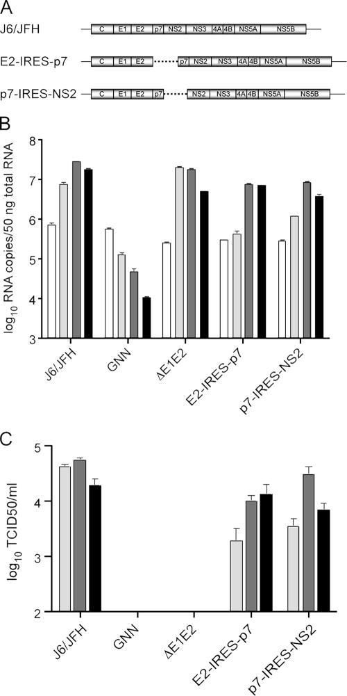
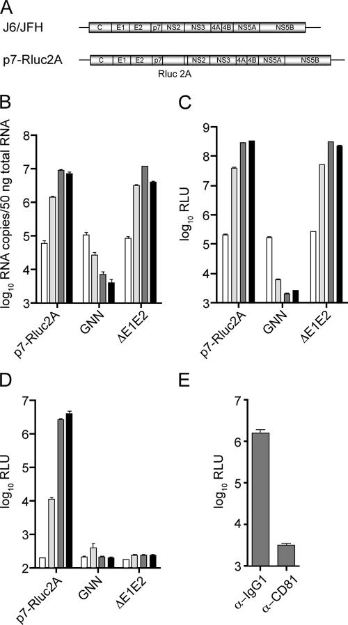
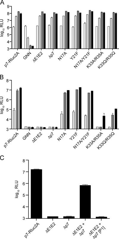
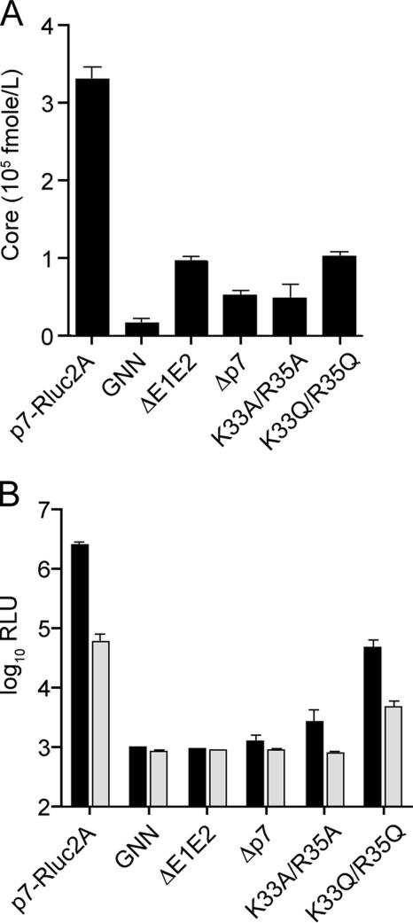
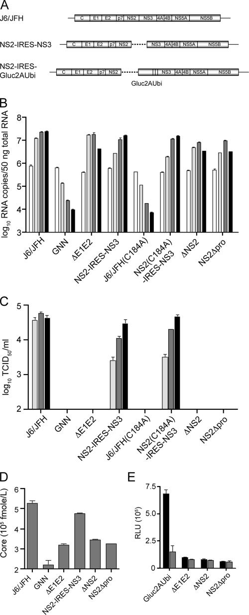
Hepatitis C virus (HCV) infection is a global health concern affecting an estimated 3% of the world's population. Recently, cell culture systems have been established, allowing recapitulation of the complete virus life cycle for the first time. Since the HCV proteins p7 and NS2 are not predicted to be major components of the virion, nor are they required for RNA replication, we investigated whether they might have other roles in the viral life cycle. Here we utilize the recently described infectious J6/JFH chimera to establish that the p7 and NS2 proteins are essential for HCV infectivity. Furthermore, unprocessed forms of p7 and NS2 were not required for this activity. Mutation of two conserved basic residues, previously shown to be important for the ion channel activity of p7 in vitro, drastically impaired infectious virus production. The protease domain of NS2 was required for infectivity, whereas its catalytic active site was dispensable. We conclude that p7 and NS2 function at an early stage of virion morphogenesis, prior to the assembly of infectious virus.


Figures

FIG. 1.
FIG. 1.
Unprocessed forms of p7 are dispensable for infectious virus production. (A) Schematic representation of full-length monocistronic genome J6/JFH (top) and bicistronic genomes E2-IRES-p7 (middle) and p7-IRES-NS2 (bottom). The dotted line indicates the location of the EMCV IRES in bicistronic genomes. (B) Replication of mono- and bicistronic genomes at 8, 24, 48, and 72 h posttransfection (white to black bars, respectively) as determined by qRT-PCR. HCV RNA copies normalized to 50 ng of total RNA. (C) Infectious virus production of mono- and bicistronic genomes determined by TCID50 assay at 24, 48, and 72 h posttransfection (light gray to black bars, respectively). The means and standard errors of the mean (SEM) of duplicate electroporations are shown. GNN, J6/JFH(GNN); ΔE1E2, J6/JFHΔE1E2.
FIG. 2.
FIG. 2.
Characterization of a novel monocistronic reporter virus. (A) Schematic representation of full-length J6/JFH (top) and the reporter virus J6/JFH(p7-Rluc2A) (bottom). The J6/JFH(p7-Rluc2A) genome contains the coding sequence for Renilla luciferase followed by the FMDV (2A) peptide between p7 and NS2. RNA replication of J6/JFH(p7-Rluc2A) as determined by qRT-PCR (B) and luciferase assay (C) at 8, 24, 48, and 72 h posttransfection (white to black bars, respectively). For qRT-PCR analysis, HCV RNA copies were normalized to 50 ng of total RNA. (D) Infectious virus production of J6/JFH(p7-Rluc2A) at 8, 24, 48, and 72 h posttransfection (white to black bars, respectively). (E) Virus neutralization of J6/JFH(p7-Rluc2A). Virus-containing supernatants were treated with the isotype control antibody, α-IgG1, or the neutralizing antibody, α-CD81. The luciferase activity was determined at 48 h postinfection. The means and SEM of at least duplicate experiments are shown. GNN, J6/JFH(p7-Rluc2A)GNN; ΔE1E2, J6/JFH(p7-Rluc2A)ΔE1E2.
FIG. 3.
FIG. 3.
p7 is essential for infectious virus production. (A) RNA replication of J6/JFH(p7-Rluc2A) genomes by luciferase assay at 8, 24, 48, and 72 h posttransfection (white to black bars, respectively). (B) Infectious virus production at 24, 48, and 72 h posttransfection (light gray to black bars, respectively). The deletions and mutations in the J6/JFH(p7-Rluc2A) genome are designated below. The positions of the point mutants are based on p7 numbering. (C) Infectious virus production at 72 h posttransfection of cells cotransfected with ΔE1E2 and Δp7 genomes (ΔE1E2+Δp7) and after one cell-free passage of the supernatant (ΔE1E2+Δp7[P1]). The means and SEM of duplicate experiments are shown. GNN, J6/JFH(p7-Rluc2A)GNN; ΔE1E2, J6/JFH(p7-Rluc2A)ΔE1E2; Δp7, Δp7-Rluc2A.
FIG. 4.
FIG. 4.
p7 functions at an early stage of virion morphogenesis. HCV core release into cell culture supernatants (A) and intracellular infectious virus accumulation (B) at 72 h posttransfection. HCV core release was determined by using a sensitive core-based ELISA (see Materials and Methods for details) on clarified cell culture supernatants harvested at 72 h posttransfection. For panel B, an analysis of infectious intracellular virus accumulation at 72 h posttransfection was performed. Lysates generated by multiple rounds of freeze-thaw were combined with isotype control antibody, α-IgGI (▪), or the HCV neutralizing antibody, α-CD81 (□), during infection of naive Huh-7.5 cells. Luciferase activity was determined at 48 h postinfection. The means and SEM of duplicate experiments are shown. GNN, J6/JFH(p7-Rluc2A)GNN; ΔE1E2, J6/JFH(p7-Rluc2A)ΔE1E2; Δp7, Δp7-Rluc2A.
FIG. 5.
FIG. 5.
NS2, but not NS2-3, is required for infectious virus production. (A) Schematic representation of full-length monocistronic J6/JFH and bicistronic NS2-IRES-NS3 genomes. A dotted line indicates location of an EMCV IRES in NS2-IRES-NS3. (B) RNA replication of mono- and bicistronic genomes by qRT-PCR at 8, 24, 48, and 72 h posttransfection (white to black bars, respectively). HCV RNA copies normalized to 50 ng of total RNA. (C) Infectious virus production of mono- and bicistronic genomes as determined by TCID50 assay at 24, 48, and 72 h posttransfection (light gray to black bars, respectively). (D) HCV core release of mono- and bicistronic genomes at 72 h posttransfection. GNN, J6/JFH(GNN); ΔE1E2, J6/JFHΔE1E2; ΔNS2, ΔNS2-IRES-NS3; NS2Δpro, NS2Δpro-IRES-NS3. (E) Intracellular infectious virus accumulation of reporter bicistronic genomes at 72 h posttransfection. Lysates generated by multiple rounds of freeze-thaw were combined with isotype control antibody, α-IgGI (▪), or the HCV neutralizing antibody, α-CD81 (□), during infection of naive Huh-7.5 cells. The luciferase activity was determined at 48 h postinfection. Gluc2AUbi, NS2-IRES-Gluc2AUbi; ΔE1E2, NS2-IRES-Gluc2AUbiΔE1E2; ΔNS2, ΔNS2-IRES-Gluc2AUbi; NS2Δpro, NS2Δpro-IRES-Gluc2AUbi. The means and SEM of duplicate experiments are shown.

References

    1. Agapov, E. V., C. L. Murray, I. Frolov, L. Qu, T. M. Myers, and C. M. Rice. 2004. Uncleaved NS2-3 is required for production of infectious bovine viral diarrhea virus. J. Virol. 78:2414-2425. - PMC - PubMed
    1. Blight, K. J., J. A. McKeating, and C. M. Rice. 2002. Highly permissive cell lines for hepatitis C virus genomic and subgenomic RNA replication. J. Virol. 76:13001-13014. - PMC - PubMed
    1. Carrere-Kremer, S., C. Montpellier, L. Lorenzo, B. Brulin, L. Cocquerel, S. Belouzard, F. Penin, and J. Dubuisson. 2004. Regulation of hepatitis C virus polyprotein processing by signal peptidase involves structural determinants at the p7 sequence junctions. J. Biol. Chem. 279:41384-41392. - PubMed
    1. Carrere-Kremer, S., C. Montpellier-Pala, L. Cocquerel, C. Wychowski, F. Penin, and J. Dubuisson. 2002. Subcellular localization and topology of the p7 polypeptide of hepatitis C virus. J. Virol. 76:3720-3730. - PMC - PubMed
    1. Clarke, D., S. Griffin, L. Beales, C. S. Gelais, S. Burgess, M. Harris, and D. Rowlands. 2006. Evidence for the formation of a heptameric ion channel complex by the hepatitis C virus p7 protein in vitro. J. Biol. Chem. 281:37057-37068. - PMC - PubMed

Publication types

LinkOut - more resources