. 2007 Sep;81(17):8944-52.
doi: 10.1128/JVI.00180-07. Epub 2007 May 30.

Natural recombination event within the capsid genomic region leading to a chimeric strain of human enterovirus B

Affiliations

Natural recombination event within the capsid genomic region leading to a chimeric strain of human enterovirus B

Lamjed Bouslama et al. J Virol. 2007 Sep.

Abstract

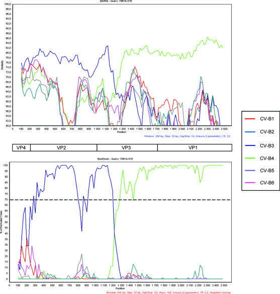
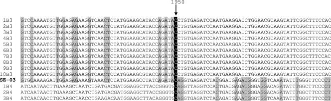
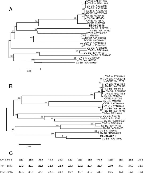
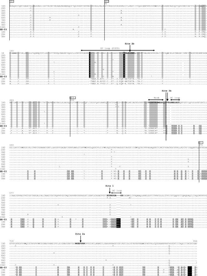
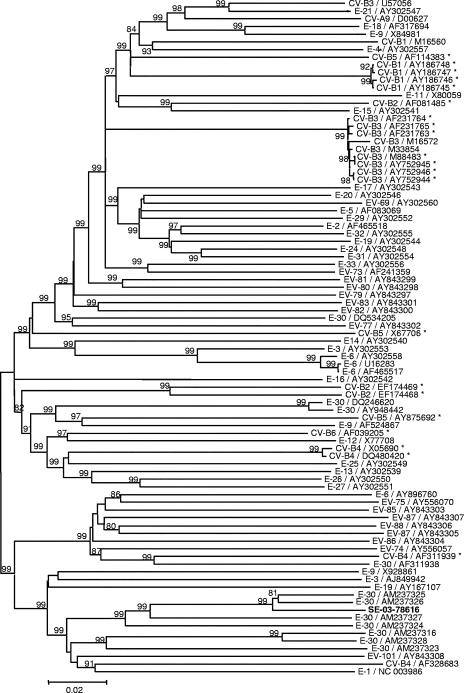
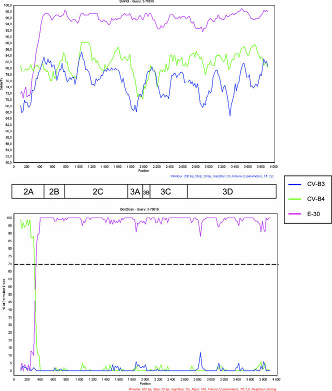
Recombination between two strains is a known phenomenon for enteroviruses replicating within a single cell. We describe a recombinant strain recovered from human stools, typed as coxsackievirus B4 (CV-B4) and CV-B3 after partial sequencing of the VP1 and VP2 coding regions, respectively. The strain was neutralized by a polyclonal CV-B3-specific antiserum but not by a CV-B4-specific antiserum. The nucleotide sequence analysis of the whole structural genomic region showed the occurrence of a recombination event at position 1950 within the VP3 capsid gene, in a region coding for the 2b antigenic site previously described for CV-B3. This observation evidences for the first time the occurrence of an interserotypic recombination within the VP2-VP3-VP1 capsid region between two nonpoliovirus enterovirus strains. The neutralization pattern suggests that the major antigenic site is located within the VP2 protein.


Figures

FIG. 1.
FIG. 1.
Similarity plot and bootscanning analysis of the P1 region comparing strain SE-03-78616 and CV-B strains with complete genomes available in databases. (Top) Similarity plot of strain SE-03-78616 versus CV-B strains constructed by the SimPlot program, version 2.5, with a sliding window of 200 nucleotides moving in 20-nucleotide steps. (Bottom) Bootscan graph of strain SE-03-78616 versus CV-B strains constructed by using the neighbor-joining tree algorithm, the Kimura-2 distance model, and 100 pseudoreplicates. The dotted line shows an arbitrary 70% reliability threshold. The following sequences (with GenBank accession numbers given in parentheses) were used: CV-B1 (AY186745 to AY186748, M16560); CV-B2 (AF081485, EF174468, EF174469); CVB-3 (AF231763 to AF231765, AY752944 to AY752946, M16572, M33854, M88483), CV-B4 (AF311939, DQ480420, X05690), CV-B5 (AF114383, AY875692, X67706), CV-B6 (AF039205).
FIG. 2.
FIG. 2.
Location of the breakpoint site of strain SE-03-78616 by nucleotide sequence alignment with the CV-B3 and CV-B4 strains listed in Fig. 1 (1B3, AY752949; 2B3, AF231765; 3B3, AF231762; 4B3, M88483; 5B3, M33854; 6B3, AY752945; 7B3, AY752944; 8B3, AF231763; 9B3, M16572; 10B3, U57056; 1B4, DQ480420; 2B4, X05690; 3B4, AF311939). Regarding positions exhibiting base homology for all tested strains of a single serotype but divergence between the two serotypes, the nucleotide shared with strain SE-03-78616 is shown against a gray background. The breakpoint site is represented by a black background.
FIG. 3.
FIG. 3.
Phylogenetic relationships in the P1 region between strain SE-03-78616 and the CV-B strains listed in Fig. 1. (A) Phylogenetic tree of the genomic region situated upstream of the breakpoint site (1,206 nucleotides, position 745 to 1950). (B) Phylogenetic tree of the genomic region situated downstream of the breakpoint site (1,357 nucleotides, position 1950 to 3306). Only bootstrap values over 75% are shown at tree nodes. (C) Nucleotide divergence (derived from the MEGA program [13] and expressed as a percentage) between strain SE-03-78616 and CV-B3/CV-B4 strains (abbreviated according to the legend to Fig. 2), upstream and downstream of the breakpoint site.
FIG. 4.
FIG. 4.
Amino acid sequence alignment of the structural proteins of strain SE-03-78616 with the CV-B3 and CV-B4 strains listed in Fig. 1 and abbreviated according to the legend to Fig. 2. Regarding positions exhibiting amino acid homology for all tested strains of a single serotype but divergence between the two serotypes, the amino acid shared with strain SE-03-78616 is shown against a gray background. Deletions or insertions having the same characteristics are shown against a black background. The solid vertical lines delineate the four structural viral proteins (VP). The dotted vertical line locates the CV-B3/CV-B4 switch on the amino acid sequence of the recombinant strain. The antigenic sites described for CV-B3 by Auvinen et al. (3) are depicted in bold type and designated according to the nomenclature of those authors. The amino acids are numbered according to the sequence of the reference strain of CV-B3 (accession number M16572). The positions of the main tertiary structures described for CV-B3 (25) are indicated by double-headed horizontal arrows above the sequences.
FIG. 5.
FIG. 5.
Phylogenetic tree showing relationships between strain SE-03-78616 and HEV-B strains in the P2-P3 region. Only bootstrap values over 75% are shown at tree nodes. The HEV-B strains are indicated by serotype and accession number in the GenBank database. Asterisks identify the strains used in Fig. 3.
FIG. 6.
FIG. 6.
Similarity plot (Top) and bootscanning analysis (Bottom) of the P2-P3 region comparing strain SE-03-78616 with CV-B3 (U57056), CV-B4 (X05690), and E-30 (AM237325) strains. The graphs were constructed as described in the legend to Fig. 1.

Similar articles

Cited by

References

    1. Altschul, S. F., W. Gish, W. Miller, E. W. Myers, and D. J. Lipman. 1990. Basic local alignment search tool. J. Mol. Biol. 215:403-410. - PubMed
    1. Andersson, P., K. Edman, and A. M. Lindberg. 2002. Molecular analysis of the echovirus 18 prototype. Evidence of interserotypic recombination with echovirus 9. Virus Res. 85:71-83. - PubMed
    1. Auvinen, P., M. J. Makela, M. Roivainen, M. Kallajoki, R. Vainionpaa, and T. Hyypia. 1993. Mapping of antigenic sites of coxsackievirus B3 by synthetic peptides. APMIS 101:517-528. - PubMed
    1. Beatrice, S. T., M. G. Katze, B. A. Zajac, and R. L. Crowell. 1980. Induction of neutralizing antibodies by the coxsackievirus B3 virion polypeptide, VP2. Virology 104:426-438. - PubMed
    1. Blomqvist, S., A. L. Bruu, M. Stenvik, and T. Hovi. 2003. Characterization of a recombinant type 3/type 2 poliovirus isolated from a healthy vaccinee and containing a chimeric capsid protein VP1. J. Gen. Virol. 84:573-580. - PubMed

Publication types

Associated data

LinkOut - more resources