. 2007 Jun 12;104(24):10128-33.
doi: 10.1073/pnas.0703735104. Epub 2007 May 30.

Structural elucidation of the m157 mouse cytomegalovirus ligand for Ly49 natural killer cell receptors

Affiliations

Structural elucidation of the m157 mouse cytomegalovirus ligand for Ly49 natural killer cell receptors

Erin J Adams et al. Proc Natl Acad Sci U S A. .

Abstract

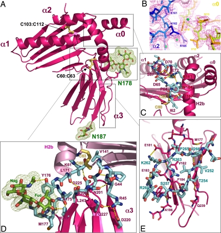
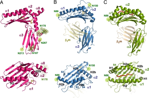
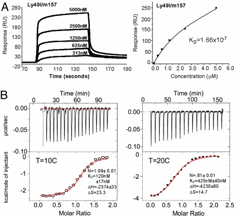
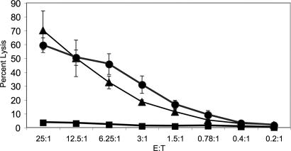
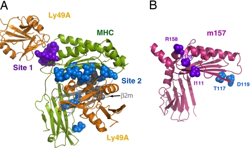
Natural killer (NK) cells express activating and inhibitory receptors that, in concert, survey cells for proper expression of cell surface major histocompatibility complex (MHC) class I molecules. The mouse cytomegalovirus encodes an MHC-like protein, m157, which is the only known viral antigen to date capable of engaging both activating (Ly49H) and inhibitory (Ly49I) NK cell receptors. We have determined the 3D structure of m157 and studied its biochemical and cellular interactions with the Ly49H and Ly49I receptors. m157 has a characteristic MHC-fold, yet possesses several unique structural features not found in other MHC class I-like molecules. m157 does not bind peptides or other small ligands, nor does it associate with beta(2)-microglobulin. Instead, m157 engages in extensive intra- and intermolecular interactions within and between its domains to generate a compact minimal MHC-like molecule. m157's binding affinity for Ly49I (K(d) approximately 0.2 microM) is significantly higher than that of classical inhibitory Ly49-MHC interactions. Analysis of viral escape mutations on m157 that render it resistant to NK killing reveals that it is likely to be recognized by Ly49H in a binding mode that differs from Ly49/MHC-I. In addition, Ly49H+ NK cells can efficiently lyse RMA cells expressing m157, despite the presence of native MHC class I. Collectively, our results show that m157 represents a structurally divergent form of MHC class I-like proteins that directly engage Ly49 receptors with appreciable affinity in a noncanonical fashion.


Conflict of interest statement

The authors declare no conflict of interest.

Figures

Fig. 1.
Fig. 1.
The 3D structure of m157. (A) Ribbon diagram of m157 showing an overall MHC class I-like structural architecture. Disulphide bonds are shown as yellow sticks, and two glycosylation sites are shown in green. (B) The experimental electron density map contoured at 1.5 ó, showing the interactions between the N-terminal α0 helix and the H2b segment of the α2 helix. (C) A network of side- and main-chain (shown in cyan) hydrogen-bonding interactions exists between the α1 and α2 domains. (D) The hinge region between the platform has extensive contacts between the α3 domain loops and the underside of the α1/α2 platform. Additional contacts are contributed by the N-linked glycosylation located at Asn-178 (shown in stick and mesh representation; green). (E) A unique C-terminal extension forms a circular loop that mediates contacts between the two β-sheets of the α3 domain's Ig sandwich (cyan-colored sticks). Hydrogen-bonding interactions are shown as dotted yellow lines, and disulfide bonds are shown in ball and stick representation (yellow).
Fig. 2.
Fig. 2.
The structural relationship of m157 with classical and nonclassical MHC class I molecules. Ribbon diagrams of the 3D structures of m157 (A), H2-T22 (B) (13), and H2-Kb (C) (35) from the side and top views, showing the altered helical and β-strand topology of m157 in relation to nonclassical and classical MHC molecules. H2-T22 and H2-Kb are shown in association with the β2m subunit [yellow (B) and orange (C)]. β-strands are numbered according to their order in the primary amino acid sequence (S1–S4) by domain, and helices are named according to their structural homology to that of the canonical MHC class I structure (12). All structural figures were generated with the program Pymol (36).
Fig. 3.
Fig. 3.
m157 binds to the inhibitory Ly49I with higher affinity than classical MHC/Ly49 interactions. (A) SPR sensogram traces (Left) and equilibrium-binding curves (Right) are shown of m157 binding to inhibitory Ly49I129 receptor. Ly49I was immobilized to a Biacore streptavidin chip via an engineered biotinylation site at the N terminus, thus ensuring appropriate orientation of the receptor on the chip surface. Concentrations of analyte (m157) are as indicated. Kd, equilibrium dissociation constant. (B) ITC traces of Ly49I and m157 at 10°C (Left) and 20°C (Right). (Upper) Rate of heat released as a function of time from injections of Ly49I into a cell containing m157. (Lower) Fitted parameters using nonlinear least-squares fitting (solid line) of the integrated areas under the respective peaks in Upper, plotted against the molar ratio of Ly49I and m157.
Fig. 4.
Fig. 4.
Expression of m157 on RMA cells overcomes class I-mediated inhibition of NK cell cytolysis. RMA tumor cells, which are protected from lysis by NK cells because of their expression of MHC class I, were transfected with m157. These m157-bearing RMA cells (filled triangles) were efficiently lysed by C57BL/6 NK cells expressing the activating Ly49H receptor. Similarly, RMA cells transfected with a ligand for the activating NKG2D receptor (filled circles) (22) were also efficiently killed by NK cells. By contrast, parental untransfected RMA cells (filled squares) were resistant to NK cell-mediated lysis because of their expression of MHC class I, consistent with prior studies. NK cell-mediated cytolysis assays were performed at various effector to target (E:T) ratios (x axis), and cytolysis was measured as percentage-specific lysis (y axis).
Fig. 5.
Fig. 5.
m157 mutants that abrogate Ly49H-mediated viral resistance do not superimpose with the canonical Ly49 binding sites on MHC class I. (A) Crystal structure of a representative Ly49–MHC complex (Ly49A/H-2Dd) (20, 24), with the residues of the MHC α-chain that involve Ly49 interaction presented in sphere style (purple, site 1; blue, site 2). (B) m157 mutations identified in previous reports, which are likely to express properly on the cell surface but fail to engage Ly49H (26, 27), were mapped onto the 3D structure (blue from ref. and purple from ref. 27). Both models are in the same orientation with respect to the platform region. Collectively, they reveal the two surface areas on m157 that are likely to involve Ly49H recognition, which are distinct from the conventional MHC–Ly49 interfaces.

References

    1. Scalzo AA, Fitzgerald NA, Wallace CR, Gibbons AE, Smart YC, Burton RC, Shellam GR. J Immunol. 1992;149:581–589. - PubMed
    1. Brown MG, Dokun AO, Heusel JW, Smith HR, Beckman DL, Blattenberger EA, Dubbelde CE, Stone LR, Scalzo AA, Yokoyama WM. Science. 2001;292:934–937. - PubMed
    1. Daniels KA, Devora G, Lai WC, O'Donnell CL, Bennett M, Welsh RM. J Exp Med. 2001;194:29–44. - PMC - PubMed
    1. Lee SH, Girard S, Macina D, Busa M, Zafer A, Belouchi A, Gros P, Vidal SM. Nat Genet. 2001;28:42–45. - PubMed
    1. Lee SH, Zafer A, de Repentigny Y, Kothary R, Tremblay ML, Gros P, Duplay P, Webb JR, Vidal SM. J Exp Med. 2003;197:515–526. - PMC - PubMed

Publication types

MeSH terms

Associated data