. 2007 Jun 19;104(25):10619-24.
doi: 10.1073/pnas.0701421104. Epub 2007 May 30.

Proline-rich tyrosine kinase 2 regulates osteoprogenitor cells and bone formation, and offers an anabolic treatment approach for osteoporosis

Affiliations

Proline-rich tyrosine kinase 2 regulates osteoprogenitor cells and bone formation, and offers an anabolic treatment approach for osteoporosis

Leonard Buckbinder et al. Proc Natl Acad Sci U S A. .

Abstract

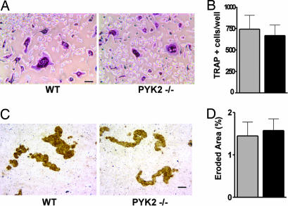
Bone is accrued and maintained primarily through the coupled actions of bone-forming osteoblasts and bone-resorbing osteoclasts. Cumulative in vitro studies indicated that proline-rich tyrosine kinase 2 (PYK2) is a positive mediator of osteoclast function and activity. However, our investigation of PYK2-/- mice did not reveal evidence supporting an essential function for PYK2 in osteoclasts either in vivo or in culture. We find that PYK2-/- mice have high bone mass resulting from an unexpected increase in bone formation. Consistent with the in vivo findings, mouse bone marrow cultures show that PYK2 deficiency enhances differentiation and activity of osteoprogenitor cells, as does expressing a PYK2-specific short hairpin RNA or dominantly interfering proteins in human mesenchymal stem cells. Furthermore, the daily administration of a small-molecule PYK2 inhibitor increases bone formation and protects against bone loss in ovariectomized rats, an established preclinical model of postmenopausal osteoporosis. In summary, we find that PYK2 regulates the differentiation of early osteoprogenitor cells across species and that inhibitors of the PYK2 have potential as a bone anabolic approach for the treatment of osteoporosis.


Conflict of interest statement

The authors declare no conflict of interest.

Figures

Fig. 1.
Fig. 1.
Bone mass and bone formation are dramatically increased in adult PYK2−/− mice at 18 weeks of age. (A) Radiography of isolated femurs from female WT control and PYK2−/− mice. (B and C) μCT images of the distal femur (B) and second lumbar vertebrae (C) of WT control and PYK2−/− mice. (D–G) Quantitative analysis of μCT images of the distal femur and second lumbar vertebrae from WT control mice and PYK2−/− mice. (D) Volumetric bone mineral density (vBMD) in WT control (hatched bars) and PYK2−/− mice (black bars). (E) Trabecular number (Tb.N). (F) Trabecular thickness (Tb.Th). (G) Cancellous bone volume per tissue volume. (H) Calcein labeling of cancellous bone in proximal tibia (green) was visualized by fluorescence micrography, with Villanueva bone counterstain (red background). (Scale bar: 100 μm.) (I–L) Dynamic histomorphometry of cancellous bone. Bone surface referent bone formation rate (BFR/BS) (I), mineralizing surface per bone surface (MS/BS) (J), and mineral apposition rate (K) are significantly increased in PYK2−/− animals. (L) Osteoclast surface (Oc.S/BS). (M) Serum CTX concentration. All data are presented as means ± SEM; n = 12–14 animals per genotype. ∗, P < 0.01; #, P < 0.05. Significance was determined by ANOVA followed by Fisher's protected least significant difference test to compare differences among groups. P < 0.05 was considered significant.
Fig. 2.
Fig. 2.
Normal osteoclastogenesis in bone marrow cultures from PYK2−/− mice. (A) Equal numbers of cells from tibial bone marrow isolated from WT control and PYK2−/− animals were plated and cultured in media supplemented with osteoprotegerin ligand and macrophage colony-stimulating factor for 4 days. Cells were fixed and stained for the presence of TRAP expression (TRAP+ cells are stained purple). (Scale bar: 50 μm.) (B) Multinucleated TRAP+ cells were counted from control (gray bar) and PYK2−/− (black bar) cultures (n = 4 wells). (C) Bone marrow-derived osteoclast cultures were grown on calcium phosphate-coated discs to assess mineral resorptive activity (osteoclast eroded surface appears brown). (Scale bar: 50 μm.) (D) Image analysis software was used to calculate the total disc eroded area in WT (gray bar) and PYK2−/− (black bar) cultures (n = 6 discs).
Fig. 3.
Fig. 3.
Osteogenesis is enhanced in bone marrow cultures from PYK2−/− mice. Equal numbers of bone marrow cells from WT (gray bars) and PYK2−/− (black bars) animals were cultured in osteogenic differentiation medium. Cell lysates were analyzed for alkaline phosphatase activity on days 14 and 19 (A) or assessed for mineralized area by quantitative fluorography of calcein labeled nodules on day 22 (B). All data are presented as means ± SEM. ∗, P < 0.01. Significance was determined by unpaired t test to compare differences between the indicated groups. P < 0.05 was considered significant.
Fig. 4.
Fig. 4.
PYK2 short hairpin RNA and dominantly interfering proteins enhance the osteogenesis of hMSC cultures. hMSCs plated in basal growth medium were infected with adenoviruses expressing a GFP control, WT PYK2, a kinase- dead PYK2 mutant (KD), a dominant interfering PYK2 splice variant (PRNK), SH, or uninfected (mock) control as indicated (see Methods and SI Fig. 7A). (A) Western blot analysis of protein extracts prepared 7 days after infection and codeveloped with monoclonal antibodies recognizing the carboxyl terminus of PYK2 and a GAPDH loading control. The positions of molecular mass markers are indicated in kilodaltons (on the right), with protein products designated on the left (NS, nonspecific). Quantitative image analysis of the full-length PYK2 doublet was normalized to GAPDH and expressed relative to PYK2 present in the GFP control sample (% GFP, at the bottom). (B) Alkaline phosphatase activity was measured on day 10. PYK2 SH was significantly different from the GFP controls (n = 3). (C) A negative well image of calcein-labeled hMSC bone nodules taken by fluorescent micrography as indicated. (D) Calcein-labeled bone nodule area was quantified by image analysis. SH, KD, and PRNK were significantly different from the GFP control (n = 3). All data are presented as means ± SEM. Significance was determined by unpaired t test to compare differences between the indicated groups. P < 0.05 was considered significant (∗, P < 0.01; #, P < 0.05).
Fig. 5.
Fig. 5.
A PYK2 inhibitor increases bone formation and blocks bone loss in ovariectomized (OVX) rats. (A) The chemical structure of PF-431396, a PYK2 inhibitor. (B) Representative three-dimensional μCT images of the distal femur from the indicated treatment groups. (C) Peripheral quantitative computed tomography (pQCT) analysis of total bone content: sham control (black bar), OVX vehicle (open bar), ethynyl estradiol (EE) (hatched bar), and PF-431396 treatment (gray bars). (D) pQCT analysis of total bone density (groups as in C). (E) Histomorphometric analysis of cancellous bone from the proximal tibia. Shown are fluorescent micrographs of the indicated treatment groups (see Table 1 for quantitative results). (F) Serum CTX levels corresponding to the indicated treatment groups (as in C). All data are presented as means ± SEM; n = 10 animals per treatment group. ∗, P < 0.05 vs. sham control; #, P < 0.05 vs. OVX; +, P < 0.05 vs. EE. Significance was determined by ANOVA followed by Fisher's protected least significant difference test to compare differences between groups. P < 0.05 was considered significant.

Comment in

  • Breaking new ground to build bone.
    Kingsley LA, Chirgwin JM, Guise TA. Kingsley LA, et al. Proc Natl Acad Sci U S A. 2007 Jun 26;104(26):10753-4. doi: 10.1073/pnas.0704357104. Epub 2007 Jun 20. Proc Natl Acad Sci U S A. 2007. PMID: 17581868 Free PMC article. Review. No abstract available.

References

    1. Schlaepfer DD, Hauck CR, Sieg DJ. Prog Biophys Mol Biol. 1999;71:435–478. - PubMed
    1. Guinamard R, Okigaki M, Schlessinger J, Ravetch JV. Nat Immunol. 2000;1:31–36. - PubMed
    1. Okigaki M, Davis C, Falasca M, Harroch S, Felsenfeld DP, Sheetz MP, Schlessinger J. Proc Natl Acad Sci USA. 2003;100:10740–10745. - PMC - PubMed
    1. Duong LT, Lakkakorpi PT, Nakamura I, Machwate M, Nagy RM, Rodan GA. J Clin Invest. 1998;102:881–892. - PMC - PubMed
    1. Lakkakorpi PT, Nakamura I, Nagy RM, Parsons JT, Rodan GA, Duong LT. J Biol Chem. 1999;274:4900–4907. - PubMed

MeSH terms