. 2007 Jun 12;104(24):10134-9.
doi: 10.1073/pnas.0700917104. Epub 2007 May 30.

Nuclear relocation of the nephrin and CD2AP-binding protein dendrin promotes apoptosis of podocytes

Affiliations

Nuclear relocation of the nephrin and CD2AP-binding protein dendrin promotes apoptosis of podocytes

Katsuhiko Asanuma et al. Proc Natl Acad Sci U S A. .

Abstract

Kidney podocytes and their slit diaphragms (SDs) form the final barrier to urinary protein loss. There is mounting evidence that SD proteins also participate in intracellular signaling pathways. The SD protein nephrin serves as a component of a signaling complex that directly links podocyte junctional integrity to actin cytoskeletal dynamics. Another SD protein, CD2-associated protein (CD2AP), is an adaptor molecule involved in podocyte homeostasis that can repress proapoptotic TGF-beta signaling in podocytes. Here we show that dendrin, a protein originally identified in telencephalic dendrites, is a constituent of the SD complex, where it directly binds to nephrin and CD2AP. In experimental glomerulonephritis, dendrin relocates from the SD to the nucleus of injured podocytes. High-dose, proapoptotic TGF-beta1 directly promotes the nuclear import of dendrin, and nuclear dendrin enhances both staurosporine- and TGF-beta1-mediated apoptosis. In summary, our results identify dendrin as an SD protein with proapoptotic signaling properties that accumulates in the podocyte nucleus in response to glomerular injury and provides a molecular target to tackle proteinuric kidney diseases. Nuclear relocation of dendrin may provide a mechanism whereby changes in SD integrity could translate into alterations of podocyte survival under pathological conditions.


Conflict of interest statement

The authors declare no conflict of interest.

Figures
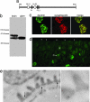
Fig. 1.
Fig. 1.
Dendrin is a component of the SD complex. (a) Dendrin contains two putative NLSs (NLS1 and NLS2), and three PPXY motifs (P1, P2, and P3). (b) Western blot analysis. In contrast to the brain, which contains both dendrin isoforms, only the 81-kDa isoform is found in isolated glomeruli. (c) Double labeling immunofluorescence microscopy of adult mouse kidney with synaptopodin confirms the expression of dendrin in podocytes. (d) During kidney development, dendrin is found in differentiating podocytes of the capillary loop stage (C) but not in undifferentiated podocytes of the S-shaped body stage (S). Arrowheads mark the kidney capsule. (e) Immunogold labeling of ultrathin frozen sections from adult mouse kidney shows the localization of dendrin in podocyte FP at the cytoplasmic insertion site of the SD (arrows).
Fig. 2.
Fig. 2.
The C terminus of dendrin interacts directly with nephrin and CD2AP. (a) Nephrin, CD2AP, and podocin from glomerular extracts (input) specifically interact with GST-dendrin but not with GST alone. (b) Coimmunoprecipitation experiments showing that endogenous dendrin interacts with nephrin in podocytes from isolated glomeruli. (Left) Immunoprecipitation (IP) with anti-dendrin. (Right) IP with anti-nephrin. No interaction is found with anti-GFP serving as negative control. (c) The GFP-tagged C-terminal (GFP-C-term) but not N-terminal (GFP-N-term) fragment of dendrin coprecipitates with FLAG-nephrin but not the FLAG control. No interaction is found between FLAG-nephrin and GFP alone. (d) GFP-C-term but not GFP-N-term also interacts with FLAG-CD2AP. No interaction was found with the GFP or FLAG controls. (e) GST-dendrin but not the GST control binds directly to FLAG-nephrin and FLAG-CD2AP, respectively.
Fig. 3.
Fig. 3.
Nuclear translocation of dendrin in experimental glomerulonephritis. (Top) In PBS-injected control mice lacking bridging podocytes, dendrin is absent from WT-1-positive podocyte nuclei. Arrowheads indicate autofluorescence of red blood cells. (Center) On day 7 after disease induction, dendrin colocalizes with WT-1 in the nucleus of a bridging podocyte that adheres to Bowman's capsule. Of note, there is also simultaneous labeling of the podocyte cell membrane (arrow). (Bottom) On day 14, in mice with active disease, dendrin can be found in the nucleus of WT-1-positive podocyte within crescents (arrows).
Fig. 4.
Fig. 4.
NLS1-mediated nuclear import of dendrin. (a) (Upper) Confocal microscopy with DAPI shows that in differentiated cultured podocytes endogenous dendrin is found in the nucleus. (Lower) Cytoplasmic dendrin is associated with intermediate filaments as shown by double labeling deconvolution microscopy with vimentin. (b) Schematic of GFP-dendrin, GFP-dendrinΔNLS1, and GFP-dendrinΔNLS2. (c) Double labeling with DAPI reveals nuclear localization of GFP-dendrin and GFP-dendrinΔNLS2. In contrast, GFP-dendrinΔNLS1 is excluded from the nucleus (arrows). (d) Similar to HEK cells, in transfected podocytes, GFP-dendrin and GFP-dendrinΔNLS2 are imported into the nucleus. In contrast, GFP-dendrinΔNLS1 is excluded from the nucleus and accumulates in the cytoplasm.
Fig. 5.
Fig. 5.
Nuclear dendrin promotes staurosporine- and TGF-β-mediated apoptosis. (a) Time course of TGF-β-induced nuclear import of dendrin after serum starvation for 24 h. Nuclear translocation of dendrin can be observed after 15 min and is completed by 30 min. No further increase is seen at 60 min. (b) Quantitative analysis of TGF-β-induced nuclear import of dendrin. For details, see Results. (c and d) Nuclear (full-length) but not cytoplasmic (ΔNLS1) dendrin enhances staurosporine- (c) and TGF-β- (d) induced apoptosis in HEK293 cells. For details, see Results. (e) Western blot analysis reveals reduction of dendrin protein expression after lentiviral knockdown of dendrin (den) mRNA expression. GAPDH shows equal protein loading. (f and g) Knockdown of dendrin expression significantly reduces staurosporine- (f) and TGF-β- (g) induced apoptosis in podocytes. For details, see Results.

References

    1. Somlo S, Mundel P. Nat Genet. 2000;24:333–335. - PubMed
    1. Kestila M, Lenkkeri U, Mannikko M, Lamerdin J, McCready P, Putaala H, Ruotsalainen V, Morita T, Nissinen M, Herva R, et al. Mol Cell. 1998;1:575–582. - PubMed
    1. Shih NY, Li J, Karpitskii V, Nguyen A, Dustin ML, Kanagawa O, Miner JH, Shaw AS. Science. 1999;286:312–315. - PubMed
    1. Kaplan JM, Kim SH, North KN, Rennke H, Correia LA, Tong HQ, Mathis BJ, Rodriguez-Perez JC, Allen PG, Beggs AH, Pollak MR. Nat Genet. 2000;24:251–256. - PubMed
    1. Boute N, Gribouval O, Roselli S, Benessy F, Lee H, Fuchshuber A, Dahan K, Gubler MC, Niaudet P, Antignac C. Nat Genet. 2000;24:349–354. - PubMed

Publication types

MeSH terms

LinkOut - more resources