

The functional impact of the intrastriatal dopamine neuron grafts in parkinsonian rats is reduced with advancing disease
- PMID: 17537955
- PMCID: PMC6672262
- DOI: 10.1523/JNEUROSCI.0626-07.2007
The functional impact of the intrastriatal dopamine neuron grafts in parkinsonian rats is reduced with advancing disease
Abstract
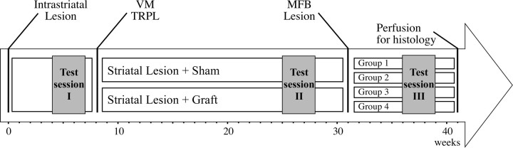
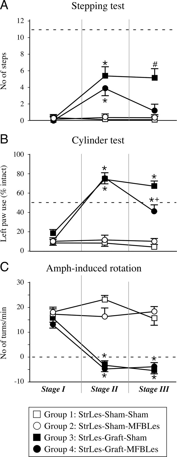
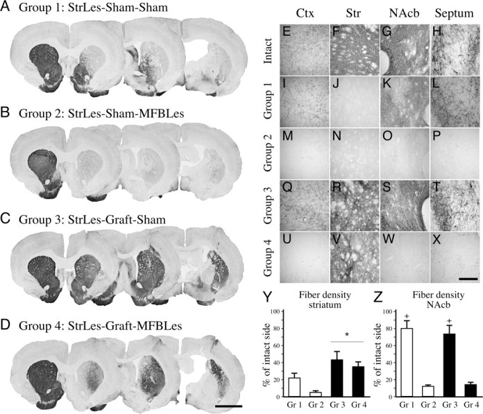
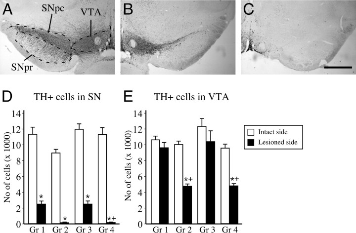
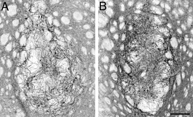
Clinical trials involving intrastriatal transplants of human embryonic mesencephalic tissue have provided proof-of-principle that nigral dopamine (DA) neurons can survive and functionally integrate into the host neural circuitry. However, the degree of graft-induced symptomatic relief differs significantly between the patients. This variability has led to investigations aimed at identifying factors that could affect the clinical outcome. The extent and pattern of dopaminergic denervation in the brain may be one of the major determinants of the functional outcome after intrastriatal DA cell grafts. Here, we report that in animals subjected to an intrastriatal 6-hydroxydopamine lesion of the striatal dopaminergic afferent, the integrity of the host dopaminergic innervation outside the areas innervated by the graft is critical for optimal function of DA neurons placed in the striatum. Established graft-induced functional recovery, as assessed in the stepping and cylinder tests, was compromised in animals in which the dopaminergic lesion was extended to include also the medial and ventral striatum as well as the cortical and limbic DA projections. Poor clinical outcome after transplantation may, thus, at least in part, be caused by dopaminergic denervation in areas outside the graft-innervated territories, and similarly beneficial effects initially observed in patients may regress if the degeneration of the host extrastriatal DA projection systems proceeds with advancing disease. This would have two implications: first, patients with advanced disease involving the ventral striatum and/or nonstriatal DA projections would be unlikely to respond well to intrastriatal DA grafts and, second, to retain the full benefit of the grafts, progression of the disease should be avoided by, for example, combining cell therapy with a neuroprotective approach.
Figures
References
-
- Brundin P, Pogarell O, Hagell P, Piccini P, Widner H, Schrag A, Kupsch A, Crabb L, Odin P, Gustavii B, Bjorklund A, Brooks DJ, Marsden CD, Oertel WH, Quinn NP, Rehncrona S, Lindvall O. Bilateral caudate and putamen grafts of embryonic mesencephalic tissue treated with lazaroids in Parkinson's disease. Brain. 2000;123:1380–1390. - PubMed
-
- Cochen V, Ribeiro MJ, Nguyen JP, Gurruchaga JM, Villafane G, Loc'h C, Defer G, Samson Y, Peschanski M, Hantraye P, Cesaro P, Remy P. Transplantation in Parkinson's disease: PET changes correlate with the amount of grafted tissue. Mov Disord. 2003;18:928–932. - PubMed
-
- Damier P, Hirsch EC, Agid Y, Graybiel AM. The substantia nigra of the human brain. II. Patterns of loss of dopamine-containing neurons in Parkinson's disease. Brain. 1999;122:1437–1448. - PubMed
-
- Dunnett SB, Robbins TW. The functional role of mesotelencephalic dopamine systems. Biol Rev Camb Philos Soc. 1992;67:491–518. - PubMed
-
- Fearnley JM, Lees AJ. Ageing and Parkinson's disease: substantia nigra regional selectivity. Brain. 1991;114:2283–2301. - PubMed
Publication types
MeSH terms
Substances
LinkOut - more resources
Full Text Sources
Other Literature Sources